

450
S. Perrin et al.
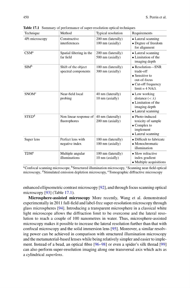
Table 17.1 Summary of performance of super-resolution optical techniques
Technique
Method
Typical resolution
Requirements
4Pi microscopy
Constructive
interferences
200 nm (laterally)
100 nm (axially)
• Lateral scanning
• Degree of freedom
for alignment
CSM a
Spatial filtering in the
far field
200 nm (laterally)
500 nm (axially)
• Lateral scanning
• Limitation of the
imaging depth
SIM b
Shift of the object
spectral components
100 nm (laterally)
300 nm (axially)
• Resolution—SNR
trade-off
• Sensitive to
out-of-focus
• Cut-off frequency
limit = 4 NA/λ
SNOM c
Near-field local
probing
40 nm (laterally)
10 nm (axially)
• Low working
distance (< λ)
• Limitation of the
imaging depth
• Lateral scanning
STED d
Non linear response of
fluorophores
40 nm (laterally)
200 nm (axially)
• Photo-induced
toxicity of sample
• Complex to
implement
• Lateral scanning
Super lens
Perfect lens with
negative index
100 nm (laterally)
100 nm (axially)
• Difficult to fabricate
• Monochromatic
illumination
TDM e
Multiple angular
illuminations
100 nm (laterally)
10 nm (axially)
• Slow refractive
index gradient
• Multiple acquisitions
a Confocal scanning microscope, b Structured illumination microscopy, c Scanning near-field optical
microscopy, d Stimulated emission depletion microscopy, e Tomographic diffractive microscopy
enhanced ellipsometric contrast microscopy [92], and through focus scanning optical
microscopy [93] (Table 17.1).
Microsphere-assisted microscopy More recently, Wang et al. demonstrated
experimentally in 2011 full-field and label-free super-resolution microscopy through
glass microspheres [94]. Introducing a transparent microsphere in a classical white
light microscope allows the diffraction limit to be overcome and the lateral resolution to reach a couple of 100 nanometres in water. Thus, microsphere-assisted
microscopy makes it possible to increase the lateral resolution further than that with
confocal microscopy and the solid immersion lens [95]. Moreover, a similar resolving power can be achieved in comparison with structured illumination microscopy
and the metamaterial-based lenses while being relatively simpler and easier to implement. Instead of a bead, an optical fibre [96–98] or even a spider’s silk thread [99]
can also perform super-resolution imaging along one transversal axis which acts as
a cylindrical superlens.
S. Perrin et al.
Table 17.1 Summary of performance of super-resolution optical techniques
Technique
Method
Typical resolution
Requirements
4Pi microscopy
Constructive
interferences
200 nm (laterally)
100 nm (axially)
• Lateral scanning
• Degree of freedom
for alignment
CSM a
Spatial filtering in the
far field
200 nm (laterally)
500 nm (axially)
• Lateral scanning
• Limitation of the
imaging depth
SIM b
Shift of the object
spectral components
100 nm (laterally)
300 nm (axially)
• Resolution—SNR
trade-off
• Sensitive to
out-of-focus
• Cut-off frequency
limit = 4 NA/λ
SNOM c
Near-field local
probing
40 nm (laterally)
10 nm (axially)
• Low working
distance (< λ)
• Limitation of the
imaging depth
• Lateral scanning
STED d
Non linear response of
fluorophores
40 nm (laterally)
200 nm (axially)
• Photo-induced
toxicity of sample
• Complex to
implement
• Lateral scanning
Super lens
Perfect lens with
negative index
100 nm (laterally)
100 nm (axially)
• Difficult to fabricate
• Monochromatic
illumination
TDM e
Multiple angular
illuminations
100 nm (laterally)
10 nm (axially)
• Slow refractive
index gradient
• Multiple acquisitions
a Confocal scanning microscope, b Structured illumination microscopy, c Scanning near-field optical
microscopy, d Stimulated emission depletion microscopy, e Tomographic diffractive microscopy
enhanced ellipsometric contrast microscopy [92], and through focus scanning optical
microscopy [93] (Table 17.1).
Microsphere-assisted microscopy More recently, Wang et al. demonstrated
experimentally in 2011 full-field and label-free super-resolution microscopy through
glass microspheres [94]. Introducing a transparent microsphere in a classical white
light microscope allows the diffraction limit to be overcome and the lateral resolution to reach a couple of 100 nanometres in water. Thus, microsphere-assisted
microscopy makes it possible to increase the lateral resolution further than that with
confocal microscopy and the solid immersion lens [95]. Moreover, a similar resolving power can be achieved in comparison with structured illumination microscopy
and the metamaterial-based lenses while being relatively simpler and easier to implement. Instead of a bead, an optical fibre [96–98] or even a spider’s silk thread [99]
can also perform super-resolution imaging along one transversal axis which acts as
a cylindrical superlens.