

16 Theoretical Foundations of Super-Resolution in Microspherical Nanoscopy
433
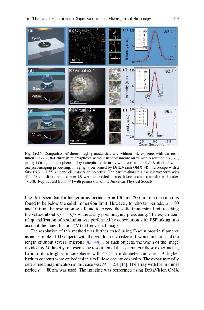
Fig. 16.16 Comparison of three imaging modalities, a–c without microspheres with the resolution ∼λ/2.2, d–f through microspheres without nanoplasmonic array with resolution ∼λ/3.7,
and g–i through microspheres using nanoplasmonic array with resolution ∼λ/6.8 obtained without post-imaging processing. Imaging is performed by DeltaVision OMX SR microscope with a
60× (NA = 1.35) silicone oil immersion objective. The barium-titanate glass microspheres with
45 − 53 µm diameters and n = 1.9 were embedded in a cellulose acetate coverslip with index
∼1.48. Reproduced from [44] with permission of the American Physical Society
line. It is seen that for longer array periods, a = 150 and 200 nm, the resolution is
found to be below the solid immersion limit. However, for shorter periods, a = 80
and 100 nm, the resolution was found to exceed the solid immersion limit reaching
the values about λ/6 − λ/7 without any post-imaging processing. The experimental quantification of resolution was performed by convolution with PSF taking into
account the magnification (M) of the virtual image.
The resolution of this method was further tested using F-actin protein filaments
as an example of 1D objects with the width on the order of few nanometers and the
length of about several microns [43, 44]. For such objects, the width of the image
divided by M directly represents the resolution of the system. For these experiments,
barium-titanate glass microspheres with 45–53 µm diameter and n = 1.9 (higher
barium content) were embedded in a cellulose acetate coverslip. The experimentally
determined magnification in this case was M = 2.4 [44]. The array with the minimal
period a = 80 nm was used. The imaging was performed using DeltaVision OMX
433
Fig. 16.16 Comparison of three imaging modalities, a–c without microspheres with the resolution ∼λ/2.2, d–f through microspheres without nanoplasmonic array with resolution ∼λ/3.7,
and g–i through microspheres using nanoplasmonic array with resolution ∼λ/6.8 obtained without post-imaging processing. Imaging is performed by DeltaVision OMX SR microscope with a
60× (NA = 1.35) silicone oil immersion objective. The barium-titanate glass microspheres with
45 − 53 µm diameters and n = 1.9 were embedded in a cellulose acetate coverslip with index
∼1.48. Reproduced from [44] with permission of the American Physical Society
line. It is seen that for longer array periods, a = 150 and 200 nm, the resolution is
found to be below the solid immersion limit. However, for shorter periods, a = 80
and 100 nm, the resolution was found to exceed the solid immersion limit reaching
the values about λ/6 − λ/7 without any post-imaging processing. The experimental quantification of resolution was performed by convolution with PSF taking into
account the magnification (M) of the virtual image.
The resolution of this method was further tested using F-actin protein filaments
as an example of 1D objects with the width on the order of few nanometers and the
length of about several microns [43, 44]. For such objects, the width of the image
divided by M directly represents the resolution of the system. For these experiments,
barium-titanate glass microspheres with 45–53 µm diameter and n = 1.9 (higher
barium content) were embedded in a cellulose acetate coverslip. The experimentally
determined magnification in this case was M = 2.4 [44]. The array with the minimal
period a = 80 nm was used. The imaging was performed using DeltaVision OMX