

16 Theoretical Foundations of Super-Resolution in Microspherical Nanoscopy
419
x/λ
y/λ
y/λ
y/λ
y/λ
-18
-15
-12
-9
-6
-3
0
3
6
-6 -3 0 3 6
0
0.95
1.9
D/λ=3.5
-6 -3 0 3 6
0
1
2
D/λ=6.735
-6 -3 0 3 6
0
0.7
1.4
D/λ=6.85
-6 -3 0 3 6
0
0.7
1.4
D/λ=9.8
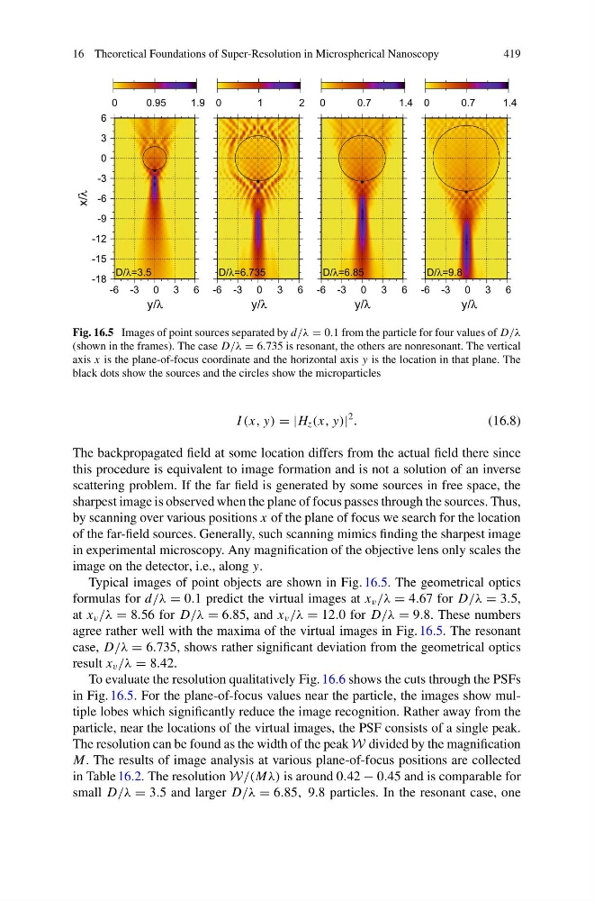
Fig. 16.5 Images of point sources separated by d/λ = 0.1 from the particle for four values of D/λ
(shown in the frames). The case D/λ = 6.735 is resonant, the others are nonresonant. The vertical
axis x is the plane-of-focus coordinate and the horizontal axis y is the location in that plane. The
black dots show the sources and the circles show the microparticles
I (x, y) = |H z (x, y)|
2
.
(16.8)
The backpropagated field at some location differs from the actual field there since
this procedure is equivalent to image formation and is not a solution of an inverse
scattering problem. If the far field is generated by some sources in free space, the
sharpest image is observed when the plane of focus passes through the sources. Thus,
by scanning over various positions x of the plane of focus we search for the location
of the far-field sources. Generally, such scanning mimics finding the sharpest image
in experimental microscopy. Any magnification of the objective lens only scales the
image on the detector, i.e., along y.
Typical images of point objects are shown in Fig. 16.5. The geometrical optics
formulas for d/λ = 0.1 predict the virtual images at x v /λ = 4.67 for D/λ = 3.5,
at x v /λ = 8.56 for D/λ = 6.85, and x v /λ = 12.0 for D/λ = 9.8. These numbers
agree rather well with the maxima of the virtual images in Fig. 16.5. The resonant
case, D/λ = 6.735, shows rather significant deviation from the geometrical optics
result x v /λ = 8.42.
To evaluate the resolution qualitatively Fig. 16.6 shows the cuts through the PSFs
in Fig. 16.5. For the plane-of-focus values near the particle, the images show multiple lobes which significantly reduce the image recognition. Rather away from the
particle, near the locations of the virtual images, the PSF consists of a single peak.
The resolution can be found as the width of the peak W divided by the magnification
M. The results of image analysis at various plane-of-focus positions are collected
in Table 16.2. The resolution W/(Mλ) is around 0.42 − 0.45 and is comparable for
small D/λ = 3.5 and larger D/λ = 6.85, 9.8 particles. In the resonant case, one
419
x/λ
y/λ
y/λ
y/λ
y/λ
-18
-15
-12
-9
-6
-3
0
3
6
-6 -3 0 3 6
0
0.95
1.9
D/λ=3.5
-6 -3 0 3 6
0
1
2
D/λ=6.735
-6 -3 0 3 6
0
0.7
1.4
D/λ=6.85
-6 -3 0 3 6
0
0.7
1.4
D/λ=9.8
Fig. 16.5 Images of point sources separated by d/λ = 0.1 from the particle for four values of D/λ
(shown in the frames). The case D/λ = 6.735 is resonant, the others are nonresonant. The vertical
axis x is the plane-of-focus coordinate and the horizontal axis y is the location in that plane. The
black dots show the sources and the circles show the microparticles
I (x, y) = |H z (x, y)|
2
.
(16.8)
The backpropagated field at some location differs from the actual field there since
this procedure is equivalent to image formation and is not a solution of an inverse
scattering problem. If the far field is generated by some sources in free space, the
sharpest image is observed when the plane of focus passes through the sources. Thus,
by scanning over various positions x of the plane of focus we search for the location
of the far-field sources. Generally, such scanning mimics finding the sharpest image
in experimental microscopy. Any magnification of the objective lens only scales the
image on the detector, i.e., along y.
Typical images of point objects are shown in Fig. 16.5. The geometrical optics
formulas for d/λ = 0.1 predict the virtual images at x v /λ = 4.67 for D/λ = 3.5,
at x v /λ = 8.56 for D/λ = 6.85, and x v /λ = 12.0 for D/λ = 9.8. These numbers
agree rather well with the maxima of the virtual images in Fig. 16.5. The resonant
case, D/λ = 6.735, shows rather significant deviation from the geometrical optics
result x v /λ = 8.42.
To evaluate the resolution qualitatively Fig. 16.6 shows the cuts through the PSFs
in Fig. 16.5. For the plane-of-focus values near the particle, the images show multiple lobes which significantly reduce the image recognition. Rather away from the
particle, near the locations of the virtual images, the PSF consists of a single peak.
The resolution can be found as the width of the peak W divided by the magnification
M. The results of image analysis at various plane-of-focus positions are collected
in Table 16.2. The resolution W/(Mλ) is around 0.42 − 0.45 and is comparable for
small D/λ = 3.5 and larger D/λ = 6.85, 9.8 particles. In the resonant case, one