

398
Z. Wang and B. Luk’yanchuk
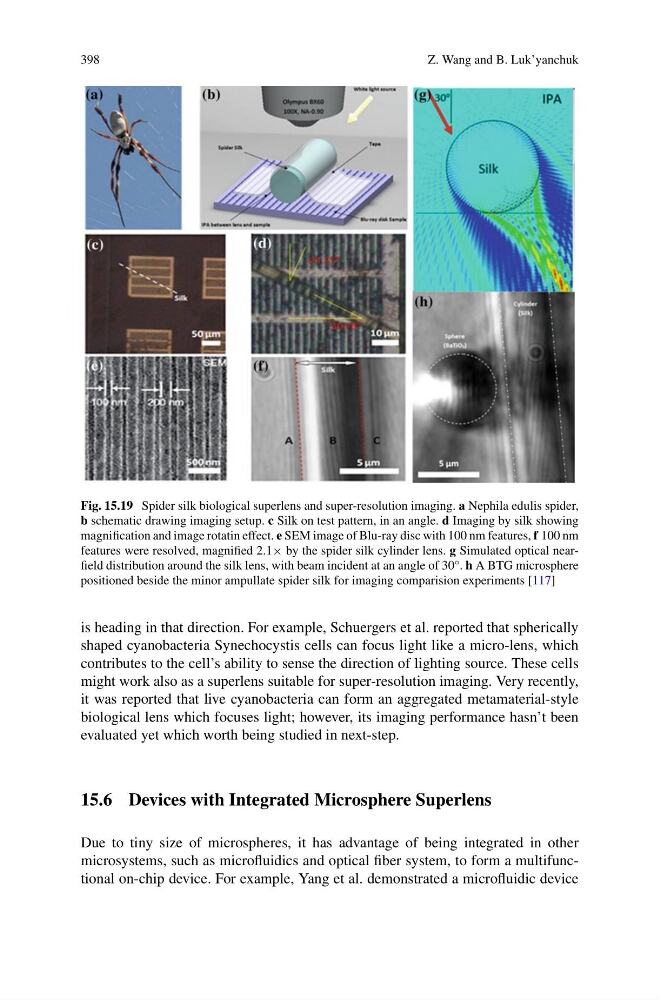
Fig. 15.19 Spider silk biological superlens and super-resolution imaging. a Nephila edulis spider,
b schematic drawing imaging setup. c Silk on test pattern, in an angle. d Imaging by silk showing
magnification and image rotatin effect. e SEM image of Blu-ray disc with 100 nm features, f 100 nm
features were resolved, magnified 2.1× by the spider silk cylinder lens. g Simulated optical nearfield distribution around the silk lens, with beam incident at an angle of 30°. h A BTG microsphere
positioned beside the minor ampullate spider silk for imaging comparision experiments [117]
is heading in that direction. For example, Schuergers et al. reported that spherically
shaped cyanobacteria Synechocystis cells can focus light like a micro-lens, which
contributes to the cell’s ability to sense the direction of lighting source. These cells
might work also as a superlens suitable for super-resolution imaging. Very recently,
it was reported that live cyanobacteria can form an aggregated metamaterial-style
biological lens which focuses light; however, its imaging performance hasn’t been
evaluated yet which worth being studied in next-step.
15.6 Devices with Integrated Microsphere Superlens
Due to tiny size of microspheres, it has advantage of being integrated in other
microsystems, such as microfluidics and optical fiber system, to form a multifunctional on-chip device. For example, Yang et al. demonstrated a microfluidic device
Z. Wang and B. Luk’yanchuk
Fig. 15.19 Spider silk biological superlens and super-resolution imaging. a Nephila edulis spider,
b schematic drawing imaging setup. c Silk on test pattern, in an angle. d Imaging by silk showing
magnification and image rotatin effect. e SEM image of Blu-ray disc with 100 nm features, f 100 nm
features were resolved, magnified 2.1× by the spider silk cylinder lens. g Simulated optical nearfield distribution around the silk lens, with beam incident at an angle of 30°. h A BTG microsphere
positioned beside the minor ampullate spider silk for imaging comparision experiments [117]
is heading in that direction. For example, Schuergers et al. reported that spherically
shaped cyanobacteria Synechocystis cells can focus light like a micro-lens, which
contributes to the cell’s ability to sense the direction of lighting source. These cells
might work also as a superlens suitable for super-resolution imaging. Very recently,
it was reported that live cyanobacteria can form an aggregated metamaterial-style
biological lens which focuses light; however, its imaging performance hasn’t been
evaluated yet which worth being studied in next-step.
15.6 Devices with Integrated Microsphere Superlens
Due to tiny size of microspheres, it has advantage of being integrated in other
microsystems, such as microfluidics and optical fiber system, to form a multifunctional on-chip device. For example, Yang et al. demonstrated a microfluidic device