

386
Z. Wang and B. Luk’yanchuk
into propagating waves, as well as the effective capture of the subwavelength information in the far field. This makes it possible to design a perfect imaging device
using dielectric nanoparticles as building block. In section below, we will show our
experimental demonstration of a fabricated TiO 2 nanoparticle-based metamaterial
solid immersion lens (mSIL) and its super-resolution imaging performance (45 nm).
15.3 Microsphere Nanoscopy: Key Progresses
In this section, we review key progress in the field, including microsphere-assisted
confocal microscopy, integrated superlensing microscope objective, scanning imaging, holography and endoscopy
15.3.1 Confocal Imaging and Resolution Quantification
In widefield, the imaging contrasts are often low and unsatisfactory due to the presence of out-of-focus light in the final image. To enhance the contrast, efforts were
required to optimize the microscope lighting and image capturing settings. In contrast, confocal microscopy generally has much better optical contrast and improved
resolution; this is achieved by placing a tiny pinhole before the detector to eliminate
the out-of-focus light in the final image. Several groups evaluated the imaging performance of microsphere-assisted confocal imaging by integrating microsphere superlens with a 405 nm laser scanning confocal microscope (e.g. Olympus OLS4000).
Wang et al. imaged 140 nm nanolines separated 40 nm away using PS particle in air,
and emphasised the importance of using single isolated particle in confocal imaging
where artefacts could arise when two neighbouring microspheres interferences with
each other [33]. Yan et al. imaged 136 nm gold nanodots separated 25 nm away,
using PS and fused silica particle in air as well [31]. Using BTG particle, Allen and
co-workers imaged 120 nm gold nanocylinders separated 60 nm away (Fig. 15.8a)
and a 185 nm nanobowtie separated 15 nm away (Fig. 15.8e) [20]. They also argued
that defining the resolution based on observation of minimal discernible features can
lead to misleading results if, for example, interpreting the 15 nm gap in nanobowtie
(Fig. 15.8e) would imply the resolution over λ/27. They suggested an approach to
compute the resolution based on the analogy with the classical theory where the
image, I(x, y), is considered as a convolution of a point spread function (PSF) and the
object’s intensity distribution function, O(u, v). This can be expressed in the standard
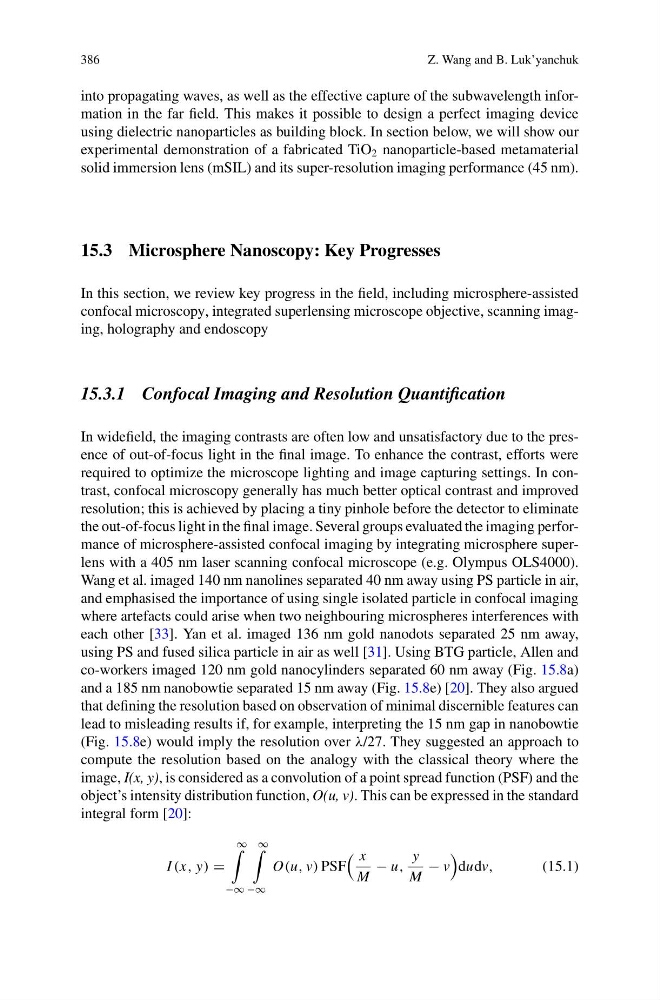
integral form [20]:
I (x, y) =
∞
−∞
∞
−∞
O(u, v) PSF
x
M
− u,
y
M
− v
dudv,
(15.1)
Z. Wang and B. Luk’yanchuk
into propagating waves, as well as the effective capture of the subwavelength information in the far field. This makes it possible to design a perfect imaging device
using dielectric nanoparticles as building block. In section below, we will show our
experimental demonstration of a fabricated TiO 2 nanoparticle-based metamaterial
solid immersion lens (mSIL) and its super-resolution imaging performance (45 nm).
15.3 Microsphere Nanoscopy: Key Progresses
In this section, we review key progress in the field, including microsphere-assisted
confocal microscopy, integrated superlensing microscope objective, scanning imaging, holography and endoscopy
15.3.1 Confocal Imaging and Resolution Quantification
In widefield, the imaging contrasts are often low and unsatisfactory due to the presence of out-of-focus light in the final image. To enhance the contrast, efforts were
required to optimize the microscope lighting and image capturing settings. In contrast, confocal microscopy generally has much better optical contrast and improved
resolution; this is achieved by placing a tiny pinhole before the detector to eliminate
the out-of-focus light in the final image. Several groups evaluated the imaging performance of microsphere-assisted confocal imaging by integrating microsphere superlens with a 405 nm laser scanning confocal microscope (e.g. Olympus OLS4000).
Wang et al. imaged 140 nm nanolines separated 40 nm away using PS particle in air,
and emphasised the importance of using single isolated particle in confocal imaging
where artefacts could arise when two neighbouring microspheres interferences with
each other [33]. Yan et al. imaged 136 nm gold nanodots separated 25 nm away,
using PS and fused silica particle in air as well [31]. Using BTG particle, Allen and
co-workers imaged 120 nm gold nanocylinders separated 60 nm away (Fig. 15.8a)
and a 185 nm nanobowtie separated 15 nm away (Fig. 15.8e) [20]. They also argued
that defining the resolution based on observation of minimal discernible features can
lead to misleading results if, for example, interpreting the 15 nm gap in nanobowtie
(Fig. 15.8e) would imply the resolution over λ/27. They suggested an approach to
compute the resolution based on the analogy with the classical theory where the
image, I(x, y), is considered as a convolution of a point spread function (PSF) and the
object’s intensity distribution function, O(u, v). This can be expressed in the standard
integral form [20]:
I (x, y) =
∞
−∞
∞
−∞
O(u, v) PSF
x
M
− u,
y
M
− v
dudv,
(15.1)