

1 Quantitative Phase Imaging: Principles and Applications
13
optics [30, 49, 78–83], medical diagnosis [84–91], and neuroscience [52, 92–97].
Due to its capabilities for label-free imaging, high sensitivity, stability, and objective
information, QPI allows investigating a sample’s structure and dynamics at broad
spatial and temporal scale [1].
1.4.1 Medical Applications
An important medical application for QPI is in pathological studies. Traditionally,
this is mostly achieved by histological methods, where one uses multiple stains to
render colors at different tissue structures and observes the sample under a conventional intensity-based microscope. The conventional methods only provide qualitative information on tissue morphology and the diagnosis outcome suffers from interobserver variability [98]. Using QPI, however, the intrinsic phase contrast accurately
maps the morphological features of sample, and more importantly, it can pick up
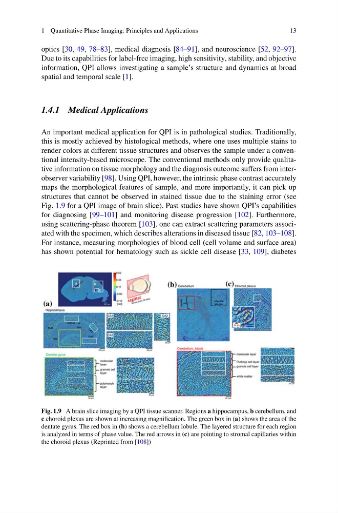
structures that cannot be observed in stained tissue due to the staining error (see
Fig. 1.9 for a QPI image of brain slice). Past studies have shown QPI’s capabilities
for diagnosing [99–101] and monitoring disease progression [102]. Furthermore,
using scattering-phase theorem [103], one can extract scattering parameters associated with the specimen, which describes alterations in diseased tissue [82, 103–108].
For instance, measuring morphologies of blood cell (cell volume and surface area)
has shown potential for hematology such as sickle cell disease [33, 109], diabetes
Fig. 1.9 A brain slice imaging by a QPI tissue scanner. Regions a hippocampus, b cerebellum, and
c choroid plexus are shown at increasing magnification. The green box in (a) shows the area of the
dentate gyrus. The red box in (b) shows a cerebellum lobule. The layered structure for each region
is analyzed in terms of phase value. The red arrows in (c) are pointing to stromal capillaries within
the choroid plexus (Reprinted from [108])
13
optics [30, 49, 78–83], medical diagnosis [84–91], and neuroscience [52, 92–97].
Due to its capabilities for label-free imaging, high sensitivity, stability, and objective
information, QPI allows investigating a sample’s structure and dynamics at broad
spatial and temporal scale [1].
1.4.1 Medical Applications
An important medical application for QPI is in pathological studies. Traditionally,
this is mostly achieved by histological methods, where one uses multiple stains to
render colors at different tissue structures and observes the sample under a conventional intensity-based microscope. The conventional methods only provide qualitative information on tissue morphology and the diagnosis outcome suffers from interobserver variability [98]. Using QPI, however, the intrinsic phase contrast accurately
maps the morphological features of sample, and more importantly, it can pick up
structures that cannot be observed in stained tissue due to the staining error (see
Fig. 1.9 for a QPI image of brain slice). Past studies have shown QPI’s capabilities
for diagnosing [99–101] and monitoring disease progression [102]. Furthermore,
using scattering-phase theorem [103], one can extract scattering parameters associated with the specimen, which describes alterations in diseased tissue [82, 103–108].
For instance, measuring morphologies of blood cell (cell volume and surface area)
has shown potential for hematology such as sickle cell disease [33, 109], diabetes
Fig. 1.9 A brain slice imaging by a QPI tissue scanner. Regions a hippocampus, b cerebellum, and
c choroid plexus are shown at increasing magnification. The green box in (a) shows the area of the
dentate gyrus. The red box in (b) shows a cerebellum lobule. The layered structure for each region
is analyzed in terms of phase value. The red arrows in (c) are pointing to stromal capillaries within
the choroid plexus (Reprinted from [108])