

13 Super-Resolution Microscopy Techniques Based …
317
operation relies on the hyperbolic dispersion law—see (13.1). In our microscopy
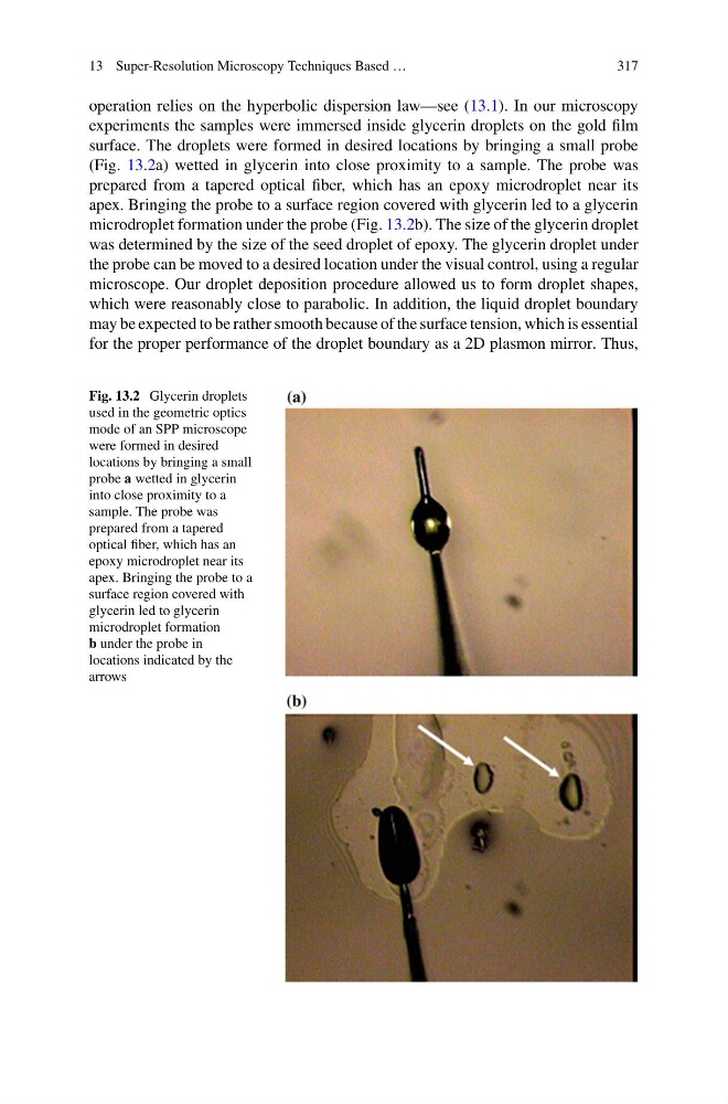
experiments the samples were immersed inside glycerin droplets on the gold film
surface. The droplets were formed in desired locations by bringing a small probe
(Fig. 13.2a) wetted in glycerin into close proximity to a sample. The probe was
prepared from a tapered optical fiber, which has an epoxy microdroplet near its
apex. Bringing the probe to a surface region covered with glycerin led to a glycerin
microdroplet formation under the probe (Fig. 13.2b). The size of the glycerin droplet
was determined by the size of the seed droplet of epoxy. The glycerin droplet under
the probe can be moved to a desired location under the visual control, using a regular
microscope. Our droplet deposition procedure allowed us to form droplet shapes,
which were reasonably close to parabolic. In addition, the liquid droplet boundary
may be expected to be rather smooth because of the surface tension, which is essential
for the proper performance of the droplet boundary as a 2D plasmon mirror. Thus,
Fig. 13.2 Glycerin droplets
used in the geometric optics
mode of an SPP microscope
were formed in desired
locations by bringing a small
probe a wetted in glycerin
into close proximity to a
sample. The probe was
prepared from a tapered
optical fiber, which has an
epoxy microdroplet near its
apex. Bringing the probe to a
surface region covered with
glycerin led to glycerin
microdroplet formation
b under the probe in
locations indicated by the
arrows
317
operation relies on the hyperbolic dispersion law—see (13.1). In our microscopy
experiments the samples were immersed inside glycerin droplets on the gold film
surface. The droplets were formed in desired locations by bringing a small probe
(Fig. 13.2a) wetted in glycerin into close proximity to a sample. The probe was
prepared from a tapered optical fiber, which has an epoxy microdroplet near its
apex. Bringing the probe to a surface region covered with glycerin led to a glycerin
microdroplet formation under the probe (Fig. 13.2b). The size of the glycerin droplet
was determined by the size of the seed droplet of epoxy. The glycerin droplet under
the probe can be moved to a desired location under the visual control, using a regular
microscope. Our droplet deposition procedure allowed us to form droplet shapes,
which were reasonably close to parabolic. In addition, the liquid droplet boundary
may be expected to be rather smooth because of the surface tension, which is essential
for the proper performance of the droplet boundary as a 2D plasmon mirror. Thus,
Fig. 13.2 Glycerin droplets
used in the geometric optics
mode of an SPP microscope
were formed in desired
locations by bringing a small
probe a wetted in glycerin
into close proximity to a
sample. The probe was
prepared from a tapered
optical fiber, which has an
epoxy microdroplet near its
apex. Bringing the probe to a
surface region covered with
glycerin led to glycerin
microdroplet formation
b under the probe in
locations indicated by the
arrows