

302
M. J. Huttunen and A. Kiviniemi
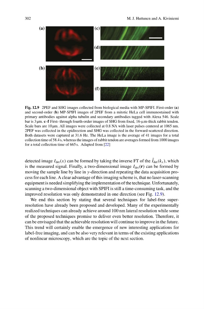
Fig. 12.9 2PEF and SHG images collected from biological media with MP-SPIFI. First-order (a)
and second-order (b) MP-SPIFI images of 2PEF from a mitotic HeLa cell immunostained with
primary antibodies against alpha tubulin and secondary antibodies tagged with Alexa 546. Scale
bar is 3 µm. c–f First- through fourth-order images of SHG from fixed, 16-μm-thick rabbit tendon.
Scale bars are 10 µm. All images were collected at 0.8 NA with laser pulses centered at 1065 nm.
2PEF was collected in the epidirection and SHG was collected in the forward-scattered direction.
Both datasets were captured at 31.6 Hz. The HeLa image is the average of 41 images for a total
collection time of 58.4 s, whereas the images of rabbit tendon are averages formed from 1000 images
for a total collection time of 665 s. Adapted from [22]
detected image I det (x) can be formed by taking the inverse FT of the ˆ
I det (k x ), which
is the measured signal. Finally, a two-dimensional image I det (r) can be formed by
moving the sample line by line in y-direction and repeating the data acquisition process for each line. A clear advantage of this imaging scheme is, that no laser-scanning
equipment is needed simplifying the implementation of the technique. Unfortunately,
scanning a two-dimensional object with SPIFI is still a time-consuming task, and the
improved resolution was only demonstrated in one direction (see Fig. 12.9).
We end this section by stating that several techniques for label-free superresolution have already been proposed and developed. Many of the experimentally
realized techniques can already achieve around 100 nm lateral resolution while some
of the proposed techniques promise to deliver even better resolution. Therefore, it
can be envisaged that the achievable resolution will continue to improve in the future.
This trend will certainly enable the emergence of new interesting applications for
label-free imaging, and can be also very relevant in terms of the existing applications
of nonlinear microscopy, which are the topic of the next section.
M. J. Huttunen and A. Kiviniemi
Fig. 12.9 2PEF and SHG images collected from biological media with MP-SPIFI. First-order (a)
and second-order (b) MP-SPIFI images of 2PEF from a mitotic HeLa cell immunostained with
primary antibodies against alpha tubulin and secondary antibodies tagged with Alexa 546. Scale
bar is 3 µm. c–f First- through fourth-order images of SHG from fixed, 16-μm-thick rabbit tendon.
Scale bars are 10 µm. All images were collected at 0.8 NA with laser pulses centered at 1065 nm.
2PEF was collected in the epidirection and SHG was collected in the forward-scattered direction.
Both datasets were captured at 31.6 Hz. The HeLa image is the average of 41 images for a total
collection time of 58.4 s, whereas the images of rabbit tendon are averages formed from 1000 images
for a total collection time of 665 s. Adapted from [22]
detected image I det (x) can be formed by taking the inverse FT of the ˆ
I det (k x ), which
is the measured signal. Finally, a two-dimensional image I det (r) can be formed by
moving the sample line by line in y-direction and repeating the data acquisition process for each line. A clear advantage of this imaging scheme is, that no laser-scanning
equipment is needed simplifying the implementation of the technique. Unfortunately,
scanning a two-dimensional object with SPIFI is still a time-consuming task, and the
improved resolution was only demonstrated in one direction (see Fig. 12.9).
We end this section by stating that several techniques for label-free superresolution have already been proposed and developed. Many of the experimentally
realized techniques can already achieve around 100 nm lateral resolution while some
of the proposed techniques promise to deliver even better resolution. Therefore, it
can be envisaged that the achievable resolution will continue to improve in the future.
This trend will certainly enable the emergence of new interesting applications for
label-free imaging, and can be also very relevant in terms of the existing applications
of nonlinear microscopy, which are the topic of the next section.