

298
M. J. Huttunen and A. Kiviniemi
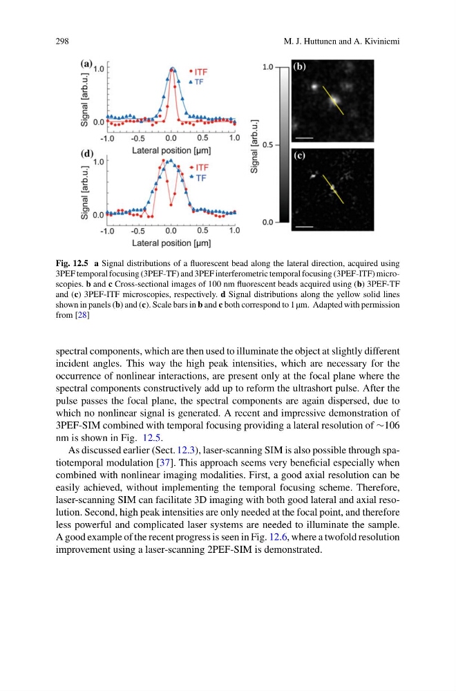
Fig. 12.5 a Signal distributions of a fluorescent bead along the lateral direction, acquired using
3PEF temporal focusing (3PEF-TF) and 3PEF interferometric temporal focusing (3PEF-ITF) microscopies. b and c Cross-sectional images of 100 nm fluorescent beads acquired using (b) 3PEF-TF
and (c) 3PEF-ITF microscopies, respectively. d Signal distributions along the yellow solid lines
shown in panels (b) and (c). Scale bars in b and c both correspond to 1 µm. Adapted with permission
from [28]
spectral components, which are then used to illuminate the object at slightly different
incident angles. This way the high peak intensities, which are necessary for the
occurrence of nonlinear interactions, are present only at the focal plane where the
spectral components constructively add up to reform the ultrashort pulse. After the
pulse passes the focal plane, the spectral components are again dispersed, due to
which no nonlinear signal is generated. A recent and impressive demonstration of
3PEF-SIM combined with temporal focusing providing a lateral resolution of ∼106
nm is shown in Fig. 12.5.
As discussed earlier (Sect. 12.3), laser-scanning SIM is also possible through spatiotemporal modulation [37]. This approach seems very beneficial especially when
combined with nonlinear imaging modalities. First, a good axial resolution can be
easily achieved, without implementing the temporal focusing scheme. Therefore,
laser-scanning SIM can facilitate 3D imaging with both good lateral and axial resolution. Second, high peak intensities are only needed at the focal point, and therefore
less powerful and complicated laser systems are needed to illuminate the sample.
A good example of the recent progress is seen in Fig. 12.6, where a twofold resolution
improvement using a laser-scanning 2PEF-SIM is demonstrated.
M. J. Huttunen and A. Kiviniemi
Fig. 12.5 a Signal distributions of a fluorescent bead along the lateral direction, acquired using
3PEF temporal focusing (3PEF-TF) and 3PEF interferometric temporal focusing (3PEF-ITF) microscopies. b and c Cross-sectional images of 100 nm fluorescent beads acquired using (b) 3PEF-TF
and (c) 3PEF-ITF microscopies, respectively. d Signal distributions along the yellow solid lines
shown in panels (b) and (c). Scale bars in b and c both correspond to 1 µm. Adapted with permission
from [28]
spectral components, which are then used to illuminate the object at slightly different
incident angles. This way the high peak intensities, which are necessary for the
occurrence of nonlinear interactions, are present only at the focal plane where the
spectral components constructively add up to reform the ultrashort pulse. After the
pulse passes the focal plane, the spectral components are again dispersed, due to
which no nonlinear signal is generated. A recent and impressive demonstration of
3PEF-SIM combined with temporal focusing providing a lateral resolution of ∼106
nm is shown in Fig. 12.5.
As discussed earlier (Sect. 12.3), laser-scanning SIM is also possible through spatiotemporal modulation [37]. This approach seems very beneficial especially when
combined with nonlinear imaging modalities. First, a good axial resolution can be
easily achieved, without implementing the temporal focusing scheme. Therefore,
laser-scanning SIM can facilitate 3D imaging with both good lateral and axial resolution. Second, high peak intensities are only needed at the focal point, and therefore
less powerful and complicated laser systems are needed to illuminate the sample.
A good example of the recent progress is seen in Fig. 12.6, where a twofold resolution
improvement using a laser-scanning 2PEF-SIM is demonstrated.