

8 Super-Resolution Imaging in Raman Microscopy
209
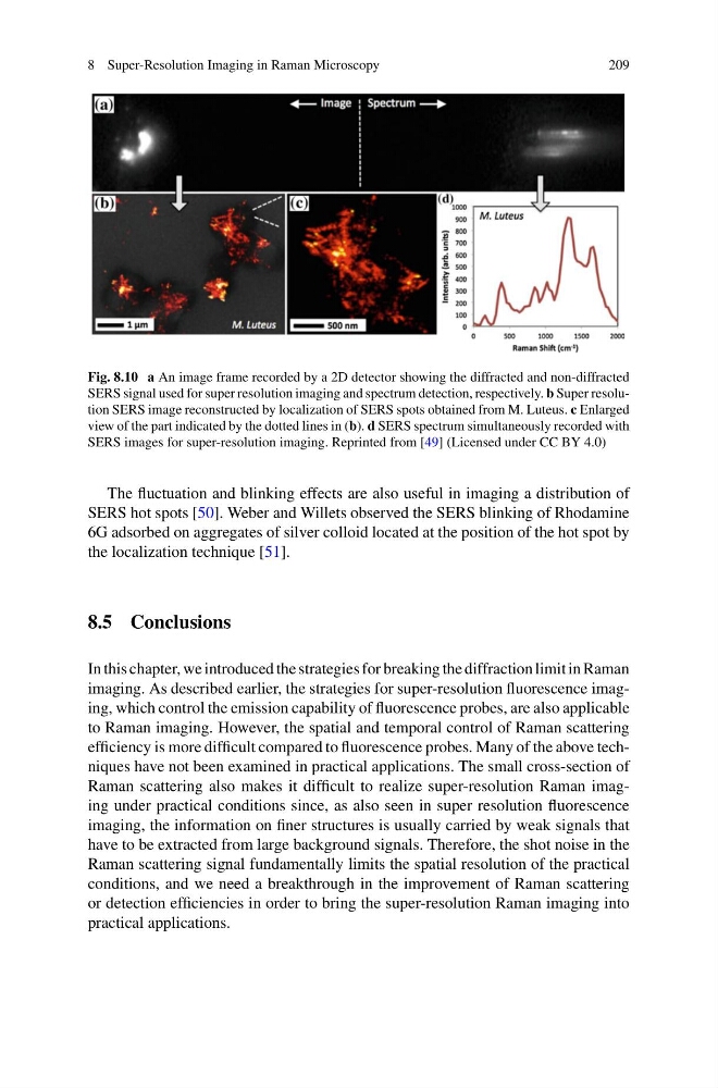
Fig. 8.10 a An image frame recorded by a 2D detector showing the diffracted and non-diffracted
SERS signal used for super resolution imaging and spectrum detection, respectively. b Super resolution SERS image reconstructed by localization of SERS spots obtained from M. Luteus. c Enlarged
view of the part indicated by the dotted lines in (b). d SERS spectrum simultaneously recorded with
SERS images for super-resolution imaging. Reprinted from [49] (Licensed under CC BY 4.0)
The fluctuation and blinking effects are also useful in imaging a distribution of
SERS hot spots [50]. Weber and Willets observed the SERS blinking of Rhodamine
6G adsorbed on aggregates of silver colloid located at the position of the hot spot by
the localization technique [51].
8.5 Conclusions
In this chapter, we introduced the strategies for breaking the diffraction limit in Raman
imaging. As described earlier, the strategies for super-resolution fluorescence imaging, which control the emission capability of fluorescence probes, are also applicable
to Raman imaging. However, the spatial and temporal control of Raman scattering
efficiency is more difficult compared to fluorescence probes. Many of the above techniques have not been examined in practical applications. The small cross-section of
Raman scattering also makes it difficult to realize super-resolution Raman imaging under practical conditions since, as also seen in super resolution fluorescence
imaging, the information on finer structures is usually carried by weak signals that
have to be extracted from large background signals. Therefore, the shot noise in the
Raman scattering signal fundamentally limits the spatial resolution of the practical
conditions, and we need a breakthrough in the improvement of Raman scattering
or detection efficiencies in order to bring the super-resolution Raman imaging into
practical applications.
209
Fig. 8.10 a An image frame recorded by a 2D detector showing the diffracted and non-diffracted
SERS signal used for super resolution imaging and spectrum detection, respectively. b Super resolution SERS image reconstructed by localization of SERS spots obtained from M. Luteus. c Enlarged
view of the part indicated by the dotted lines in (b). d SERS spectrum simultaneously recorded with
SERS images for super-resolution imaging. Reprinted from [49] (Licensed under CC BY 4.0)
The fluctuation and blinking effects are also useful in imaging a distribution of
SERS hot spots [50]. Weber and Willets observed the SERS blinking of Rhodamine
6G adsorbed on aggregates of silver colloid located at the position of the hot spot by
the localization technique [51].
8.5 Conclusions
In this chapter, we introduced the strategies for breaking the diffraction limit in Raman
imaging. As described earlier, the strategies for super-resolution fluorescence imaging, which control the emission capability of fluorescence probes, are also applicable
to Raman imaging. However, the spatial and temporal control of Raman scattering
efficiency is more difficult compared to fluorescence probes. Many of the above techniques have not been examined in practical applications. The small cross-section of
Raman scattering also makes it difficult to realize super-resolution Raman imaging under practical conditions since, as also seen in super resolution fluorescence
imaging, the information on finer structures is usually carried by weak signals that
have to be extracted from large background signals. Therefore, the shot noise in the
Raman scattering signal fundamentally limits the spatial resolution of the practical
conditions, and we need a breakthrough in the improvement of Raman scattering
or detection efficiencies in order to bring the super-resolution Raman imaging into
practical applications.