

7 Label-Free Pump–Probe Nanoscopy
187
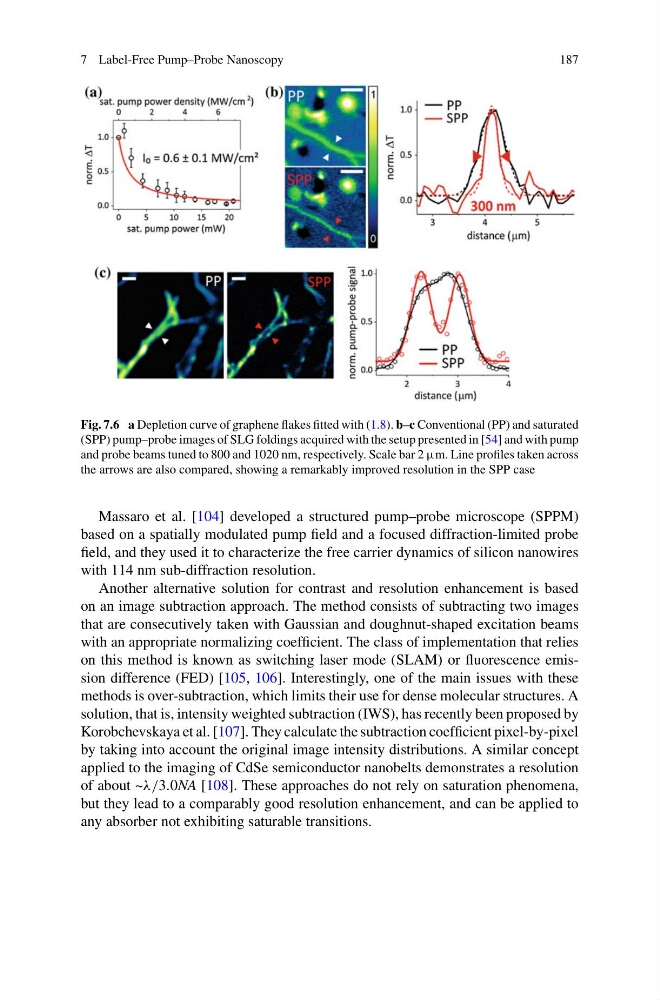
Fig. 7.6 a Depletion curve of graphene flakes fitted with (1.8). b–c Conventional (PP) and saturated
(SPP) pump–probe images of SLG foldings acquired with the setup presented in [54] and with pump
and probe beams tuned to 800 and 1020 nm, respectively. Scale bar 2 μm. Line profiles taken across
the arrows are also compared, showing a remarkably improved resolution in the SPP case
Massaro et al. [104] developed a structured pump–probe microscope (SPPM)
based on a spatially modulated pump field and a focused diffraction-limited probe
field, and they used it to characterize the free carrier dynamics of silicon nanowires
with 114 nm sub-diffraction resolution.
Another alternative solution for contrast and resolution enhancement is based
on an image subtraction approach. The method consists of subtracting two images
that are consecutively taken with Gaussian and doughnut-shaped excitation beams
with an appropriate normalizing coefficient. The class of implementation that relies
on this method is known as switching laser mode (SLAM) or fluorescence emission difference (FED) [105, 106]. Interestingly, one of the main issues with these
methods is over-subtraction, which limits their use for dense molecular structures. A
solution, that is, intensity weighted subtraction (IWS), has recently been proposed by
Korobchevskaya et al. [107]. They calculate the subtraction coefficient pixel-by-pixel
by taking into account the original image intensity distributions. A similar concept
applied to the imaging of CdSe semiconductor nanobelts demonstrates a resolution
of about ~λ/3.0NA [108]. These approaches do not rely on saturation phenomena,
but they lead to a comparably good resolution enhancement, and can be applied to
any absorber not exhibiting saturable transitions.
187
Fig. 7.6 a Depletion curve of graphene flakes fitted with (1.8). b–c Conventional (PP) and saturated
(SPP) pump–probe images of SLG foldings acquired with the setup presented in [54] and with pump
and probe beams tuned to 800 and 1020 nm, respectively. Scale bar 2 μm. Line profiles taken across
the arrows are also compared, showing a remarkably improved resolution in the SPP case
Massaro et al. [104] developed a structured pump–probe microscope (SPPM)
based on a spatially modulated pump field and a focused diffraction-limited probe
field, and they used it to characterize the free carrier dynamics of silicon nanowires
with 114 nm sub-diffraction resolution.
Another alternative solution for contrast and resolution enhancement is based
on an image subtraction approach. The method consists of subtracting two images
that are consecutively taken with Gaussian and doughnut-shaped excitation beams
with an appropriate normalizing coefficient. The class of implementation that relies
on this method is known as switching laser mode (SLAM) or fluorescence emission difference (FED) [105, 106]. Interestingly, one of the main issues with these
methods is over-subtraction, which limits their use for dense molecular structures. A
solution, that is, intensity weighted subtraction (IWS), has recently been proposed by
Korobchevskaya et al. [107]. They calculate the subtraction coefficient pixel-by-pixel
by taking into account the original image intensity distributions. A similar concept
applied to the imaging of CdSe semiconductor nanobelts demonstrates a resolution
of about ~λ/3.0NA [108]. These approaches do not rely on saturation phenomena,
but they lead to a comparably good resolution enhancement, and can be applied to
any absorber not exhibiting saturable transitions.