

182
P. Bianchini et al.
flow [72, 73], and its excited state lifetime turned out to be a useful indicator of the
oxygenation level [13].
Apart from biological pigments studies, pump–probe microscopy is also used in
identifying historical art pigments and/or pigment mixtures, to study their 3D-layer
structure and their method of application [74, 75].
7.3 Breaking the Diffraction Limit in Pump–Probe
Microscopy
Diffraction is the basis of image formation in an optical microscope, and the cause
of its limited resolving power. The image of a point source through an objective
lens does not look like an infinitely small point in the image plane, but instead, a
constructive and destructive interference produces a characteristic diffraction pattern
which consists of a central bright spot (Airy disk) surrounded by concentric rings
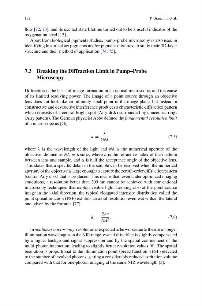
(Airy pattern). The German physicist Abbe defined the fundamental resolution limit
of a microscope as [76]
d =
λ
2NA
,
(7.5)
where λ is the wavelength of the light and NA is the numerical aperture of the
objective, defined as NA = n sin α, where n is the refractive index of the medium
between lens and sample, and α is half the acceptance angle of the objective lens.
This states that a specific detail in the sample can be resolved when the numerical
aperture of the objective is large enough to capture the zeroth-order diffraction pattern
(central Airy disk) that is produced. This means that, even under optimized imaging
conditions, a resolution better than 200 nm cannot be achieved with conventional
microscopy techniques that exploit visible light. Looking also at the point source
image in the axial direction, the typical elongated intensity distribution called the
point spread function (PSF) exhibits an axial resolution even worse than the lateral
one, given by the formula [77]:
d z =
2λn
NA 2 .
(7.6)
In nonlinear microscopy, resolution is expected to be worse due to the use of longer
illumination wavelengths in the NIR range, even if this effect is slightly compensated
by a higher background signal suppression and by the spatial confinement of the
multi-photon interaction, leading to slightly better resolution values [6]. The spatial
resolution is proportional to the illumination point spread function (IPSF) elevated
to the number of involved photons, getting a considerably reduced excitation volume
compared with that for one-photon imaging at the same NIR wavelength [3].
P. Bianchini et al.
flow [72, 73], and its excited state lifetime turned out to be a useful indicator of the
oxygenation level [13].
Apart from biological pigments studies, pump–probe microscopy is also used in
identifying historical art pigments and/or pigment mixtures, to study their 3D-layer
structure and their method of application [74, 75].
7.3 Breaking the Diffraction Limit in Pump–Probe
Microscopy
Diffraction is the basis of image formation in an optical microscope, and the cause
of its limited resolving power. The image of a point source through an objective
lens does not look like an infinitely small point in the image plane, but instead, a
constructive and destructive interference produces a characteristic diffraction pattern
which consists of a central bright spot (Airy disk) surrounded by concentric rings
(Airy pattern). The German physicist Abbe defined the fundamental resolution limit
of a microscope as [76]
d =
λ
2NA
,
(7.5)
where λ is the wavelength of the light and NA is the numerical aperture of the
objective, defined as NA = n sin α, where n is the refractive index of the medium
between lens and sample, and α is half the acceptance angle of the objective lens.
This states that a specific detail in the sample can be resolved when the numerical
aperture of the objective is large enough to capture the zeroth-order diffraction pattern
(central Airy disk) that is produced. This means that, even under optimized imaging
conditions, a resolution better than 200 nm cannot be achieved with conventional
microscopy techniques that exploit visible light. Looking also at the point source
image in the axial direction, the typical elongated intensity distribution called the
point spread function (PSF) exhibits an axial resolution even worse than the lateral
one, given by the formula [77]:
d z =
2λn
NA 2 .
(7.6)
In nonlinear microscopy, resolution is expected to be worse due to the use of longer
illumination wavelengths in the NIR range, even if this effect is slightly compensated
by a higher background signal suppression and by the spatial confinement of the
multi-photon interaction, leading to slightly better resolution values [6]. The spatial
resolution is proportional to the illumination point spread function (IPSF) elevated
to the number of involved photons, getting a considerably reduced excitation volume
compared with that for one-photon imaging at the same NIR wavelength [3].