

142
C. Li and J.-X. Cheng
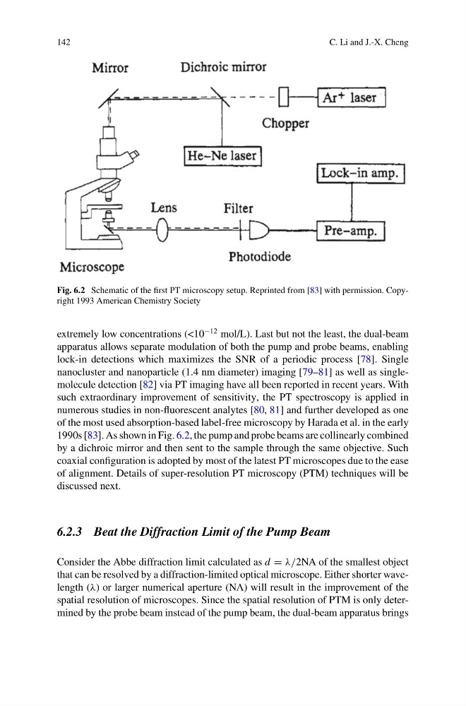
Fig. 6.2 Schematic of the first PT microscopy setup. Reprinted from [83] with permission. Copyright 1993 American Chemistry Society
extremely low concentrations (<10
−12 mol/L). Last but not the least, the dual-beam
apparatus allows separate modulation of both the pump and probe beams, enabling
lock-in detections which maximizes the SNR of a periodic process [78]. Single
nanocluster and nanoparticle (1.4 nm diameter) imaging [79–81] as well as singlemolecule detection [82] via PT imaging have all been reported in recent years. With
such extraordinary improvement of sensitivity, the PT spectroscopy is applied in
numerous studies in non-fluorescent analytes [80, 81] and further developed as one
of the most used absorption-based label-free microscopy by Harada et al. in the early
1990s [83]. As shown in Fig. 6.2, the pump and probe beams are collinearly combined
by a dichroic mirror and then sent to the sample through the same objective. Such
coaxial configuration is adopted by most of the latest PT microscopes due to the ease
of alignment. Details of super-resolution PT microscopy (PTM) techniques will be
discussed next.
6.2.3 Beat the Diffraction Limit of the Pump Beam
Consider the Abbe diffraction limit calculated as d = λ/2NA of the smallest object
that can be resolved by a diffraction-limited optical microscope. Either shorter wavelength (λ) or larger numerical aperture (NA) will result in the improvement of the
spatial resolution of microscopes. Since the spatial resolution of PTM is only determined by the probe beam instead of the pump beam, the dual-beam apparatus brings
C. Li and J.-X. Cheng
Fig. 6.2 Schematic of the first PT microscopy setup. Reprinted from [83] with permission. Copyright 1993 American Chemistry Society
extremely low concentrations (<10
−12 mol/L). Last but not the least, the dual-beam
apparatus allows separate modulation of both the pump and probe beams, enabling
lock-in detections which maximizes the SNR of a periodic process [78]. Single
nanocluster and nanoparticle (1.4 nm diameter) imaging [79–81] as well as singlemolecule detection [82] via PT imaging have all been reported in recent years. With
such extraordinary improvement of sensitivity, the PT spectroscopy is applied in
numerous studies in non-fluorescent analytes [80, 81] and further developed as one
of the most used absorption-based label-free microscopy by Harada et al. in the early
1990s [83]. As shown in Fig. 6.2, the pump and probe beams are collinearly combined
by a dichroic mirror and then sent to the sample through the same objective. Such
coaxial configuration is adopted by most of the latest PT microscopes due to the ease
of alignment. Details of super-resolution PT microscopy (PTM) techniques will be
discussed next.
6.2.3 Beat the Diffraction Limit of the Pump Beam
Consider the Abbe diffraction limit calculated as d = λ/2NA of the smallest object
that can be resolved by a diffraction-limited optical microscope. Either shorter wavelength (λ) or larger numerical aperture (NA) will result in the improvement of the
spatial resolution of microscopes. Since the spatial resolution of PTM is only determined by the probe beam instead of the pump beam, the dual-beam apparatus brings