

4 Tomographic Diffractive Microscopy …
101
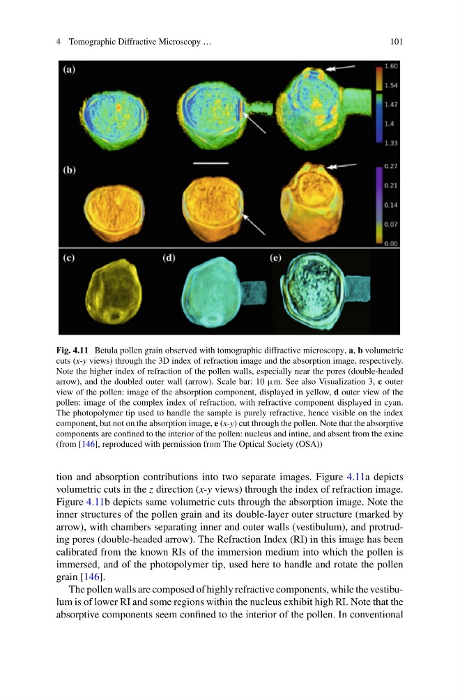
Fig. 4.11 Betula pollen grain observed with tomographic diffractive microscopy, a, b volumetric
cuts (x-y views) through the 3D index of refraction image and the absorption image, respectively.
Note the higher index of refraction of the pollen walls, especially near the pores (double-headed
arrow), and the doubled outer wall (arrow). Scale bar: 10 μm. See also Visualization 3, c outer
view of the pollen: image of the absorption component, displayed in yellow, d outer view of the
pollen: image of the complex index of refraction, with refractive component displayed in cyan.
The photopolymer tip used to handle the sample is purely refractive, hence visible on the index
component, but not on the absorption image, e (x-y) cut through the pollen. Note that the absorptive
components are confined to the interior of the pollen: nucleus and intine, and absent from the exine
(from [146], reproduced with permission from The Optical Society (OSA))
tion and absorption contributions into two separate images. Figure 4.11a depicts
volumetric cuts in the z direction (x-y views) through the index of refraction image.
Figure 4.11b depicts same volumetric cuts through the absorption image. Note the
inner structures of the pollen grain and its double-layer outer structure (marked by
arrow), with chambers separating inner and outer walls (vestibulum), and protruding pores (double-headed arrow). The Refraction Index (RI) in this image has been
calibrated from the known RIs of the immersion medium into which the pollen is
immersed, and of the photopolymer tip, used here to handle and rotate the pollen
grain [146].
The pollen walls are composed of highly refractive components, while the vestibulum is of lower RI and some regions within the nucleus exhibit high RI. Note that the
absorptive components seem confined to the interior of the pollen. In conventional
101
Fig. 4.11 Betula pollen grain observed with tomographic diffractive microscopy, a, b volumetric
cuts (x-y views) through the 3D index of refraction image and the absorption image, respectively.
Note the higher index of refraction of the pollen walls, especially near the pores (double-headed
arrow), and the doubled outer wall (arrow). Scale bar: 10 μm. See also Visualization 3, c outer
view of the pollen: image of the absorption component, displayed in yellow, d outer view of the
pollen: image of the complex index of refraction, with refractive component displayed in cyan.
The photopolymer tip used to handle the sample is purely refractive, hence visible on the index
component, but not on the absorption image, e (x-y) cut through the pollen. Note that the absorptive
components are confined to the interior of the pollen: nucleus and intine, and absent from the exine
(from [146], reproduced with permission from The Optical Society (OSA))
tion and absorption contributions into two separate images. Figure 4.11a depicts
volumetric cuts in the z direction (x-y views) through the index of refraction image.
Figure 4.11b depicts same volumetric cuts through the absorption image. Note the
inner structures of the pollen grain and its double-layer outer structure (marked by
arrow), with chambers separating inner and outer walls (vestibulum), and protruding pores (double-headed arrow). The Refraction Index (RI) in this image has been
calibrated from the known RIs of the immersion medium into which the pollen is
immersed, and of the photopolymer tip, used here to handle and rotate the pollen
grain [146].
The pollen walls are composed of highly refractive components, while the vestibulum is of lower RI and some regions within the nucleus exhibit high RI. Note that the
absorptive components seem confined to the interior of the pollen. In conventional