

2 Human Brain Anatomy in 3D
39
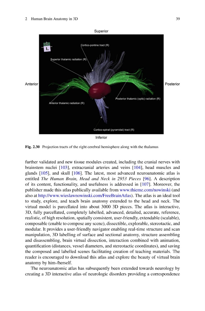
Fig. 2.30 Projection tracts of the right cerebral hemisphere along with the thalamus
further validated and new tissue modules created, including the cranial nerves with
brainstem nuclei [103], extracranial arteries and veins [104], head muscles and
glands [105], and skull [106]. The latest, most advanced neuroanatomic atlas is
entitled The Human Brain, Head and Neck in 2953 Pieces [96]. A description
of its content, functionality, and usefulness is addressed in [107]. Moreover, the
publisher made this atlas publically available from www.thieme.com/nowinski (and
also at http://www.wieslawnowinski.com/FreeBrainAtlas). The atlas is an ideal tool
to study, explore, and teach brain anatomy extended to the head and neck. The
virtual model is parcellated into about 3000 3D pieces. The atlas is interactive,
3D, fully parcellated, completely labelled, advanced, detailed, accurate, reference,
realistic, of high resolution, spatially consistent, user-friendly, extendable (scalable),
composable (enable to compose any scene), dissectible, explorable, stereotactic, and
modular. It provides a user-friendly navigator enabling real-time structure and scan
manipulation, 3D labelling of surface and sectional anatomy, structure assembling
and disassembling, brain virtual dissection, interaction combined with animation,
quantification (distances, vessel diameters, and stereotactic coordinates), and saving
the composed and labelled scenes facilitating creation of teaching materials. The
reader is encouraged to download this atlas and explore the beauty of virtual brain
anatomy by him-/herself.
The neuroanatomic atlas has subsequently been extended towards neurology by
creating a 3D interactive atlas of neurologic disorders providing a correspondence
39
Fig. 2.30 Projection tracts of the right cerebral hemisphere along with the thalamus
further validated and new tissue modules created, including the cranial nerves with
brainstem nuclei [103], extracranial arteries and veins [104], head muscles and
glands [105], and skull [106]. The latest, most advanced neuroanatomic atlas is
entitled The Human Brain, Head and Neck in 2953 Pieces [96]. A description
of its content, functionality, and usefulness is addressed in [107]. Moreover, the
publisher made this atlas publically available from www.thieme.com/nowinski (and
also at http://www.wieslawnowinski.com/FreeBrainAtlas). The atlas is an ideal tool
to study, explore, and teach brain anatomy extended to the head and neck. The
virtual model is parcellated into about 3000 3D pieces. The atlas is interactive,
3D, fully parcellated, completely labelled, advanced, detailed, accurate, reference,
realistic, of high resolution, spatially consistent, user-friendly, extendable (scalable),
composable (enable to compose any scene), dissectible, explorable, stereotactic, and
modular. It provides a user-friendly navigator enabling real-time structure and scan
manipulation, 3D labelling of surface and sectional anatomy, structure assembling
and disassembling, brain virtual dissection, interaction combined with animation,
quantification (distances, vessel diameters, and stereotactic coordinates), and saving
the composed and labelled scenes facilitating creation of teaching materials. The
reader is encouraged to download this atlas and explore the beauty of virtual brain
anatomy by him-/herself.
The neuroanatomic atlas has subsequently been extended towards neurology by
creating a 3D interactive atlas of neurologic disorders providing a correspondence