

13 Computational Biomechanics of the Brain in the Operating Theatre
331
In the opposite, constraint forces λ col applied on the collision models are weighted
with the same barycentric coefficients and converted in equivalent nodal forces on
the finite element mesh λ FE :
λ FE = J
T λ col
(13.3)
13.3.2 Intra-operative Datasets
Intra-operatively, registration data are extracted from the navigated ultrasound
images. Power Doppler and B-mode signals are recorded simultaneously, and the
corresponding 3D image volumes are reconstructed. In a clinical context, the set of
algorithms presented in the following paragraphs will have to be performed during
the surgical procedure. They must therefore be fast, a few seconds, and almost fully
automatic to allow their use in a clinical process.
13.3.2.1 Vascular Tree from Power Doppler Ultrasound Images
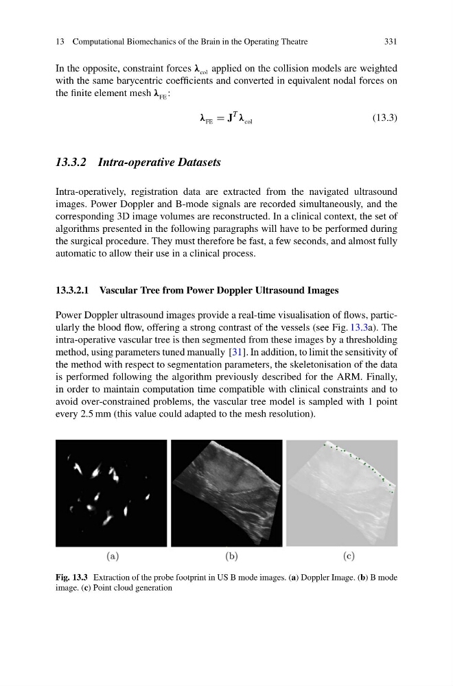
Power Doppler ultrasound images provide a real-time visualisation of flows, particularly the blood flow, offering a strong contrast of the vessels (see Fig. 13.3a). The
intra-operative vascular tree is then segmented from these images by a thresholding
method, using parameters tuned manually [31]. In addition, to limit the sensitivity of
the method with respect to segmentation parameters, the skeletonisation of the data
is performed following the algorithm previously described for the ARM. Finally,
in order to maintain computation time compatible with clinical constraints and to
avoid over-constrained problems, the vascular tree model is sampled with 1 point
every 2.5 mm (this value could adapted to the mesh resolution).
Fig. 13.3 Extraction of the probe footprint in US B mode images. (a) Doppler Image. (b) B mode
image. (c) Point cloud generation
331
In the opposite, constraint forces λ col applied on the collision models are weighted
with the same barycentric coefficients and converted in equivalent nodal forces on
the finite element mesh λ FE :
λ FE = J
T λ col
(13.3)
13.3.2 Intra-operative Datasets
Intra-operatively, registration data are extracted from the navigated ultrasound
images. Power Doppler and B-mode signals are recorded simultaneously, and the
corresponding 3D image volumes are reconstructed. In a clinical context, the set of
algorithms presented in the following paragraphs will have to be performed during
the surgical procedure. They must therefore be fast, a few seconds, and almost fully
automatic to allow their use in a clinical process.
13.3.2.1 Vascular Tree from Power Doppler Ultrasound Images
Power Doppler ultrasound images provide a real-time visualisation of flows, particularly the blood flow, offering a strong contrast of the vessels (see Fig. 13.3a). The
intra-operative vascular tree is then segmented from these images by a thresholding
method, using parameters tuned manually [31]. In addition, to limit the sensitivity of
the method with respect to segmentation parameters, the skeletonisation of the data
is performed following the algorithm previously described for the ARM. Finally,
in order to maintain computation time compatible with clinical constraints and to
avoid over-constrained problems, the vascular tree model is sampled with 1 point
every 2.5 mm (this value could adapted to the mesh resolution).
Fig. 13.3 Extraction of the probe footprint in US B mode images. (a) Doppler Image. (b) B mode
image. (c) Point cloud generation