

2 Human Brain Anatomy in 3D
25
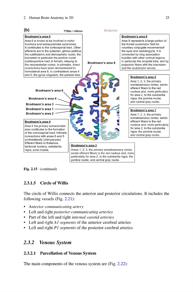
Brodmann’s area 8
Area 8 represents a large portion of
the frontal oculomotor field for
voluntary conjugate movementsof
the eyes and cephalogyria. It is
connected by long association
bundles with other cortical regions,
in particular the occipital lobe, and by
projection fibers with the brainstem
and the oculomotor nerves.
Brodmann’s area 8
TT88a / +40mm
(b)
Anterior
Brodmann’s area 6
Area 6 is known to be involved in motor
functions and extrapyramidal syndromes.
It contributes to the corticospinal tract. Other
efferents are to the putamen, globus pallidus,
the subthalamic and diencephalic nuclei, the
brainstem in particular the pontine nuclei
(corticopontine tract of Arnod), relaying to
the neocerebellar cortex. In primates, direct
connections have been demonstrated to
homolateral area 8, to contralateral areas 4
and 6, the gyrus cingularis, the parietal lobe.
Brodmann’s area 6
Brodmann’s area 4
Area 4 the primary somatomotor
area contibutes to the formation
of the cotricospinal tract. Intimate
connections with areas 6 and 5
contralaterally (chimpanzee).
Efferent fibers to thalamus,
lenticular nucleus, substantia
nigra, zona incerta.
Brodmann’s area 4
Brodmann’s area 3
Areas 1, 2, 3, the primary somatosensory cortex,
sends efferent fibers to the red nucleus and, more
particularly for area 2, to the substantia nigra, the
pontine nuclei, and central gray nuclei.
Brodmann’s area 3
Brodmann’s area 1
Area 1, 2, 3, the primary
somatosensory cortex, sends
efferent fibers to the red
nucleus and, more particularly
for area 2, to the substantia
nigra, the pontine nuclei,
and central gray nuclei.
Brodmann’s area 1
Brodmann’s area 2
Area 1, 2, 3, the primary
somatosensory cortex, sends
efferent fibers to the red
nucleus and, more particulary
for area 2, to the substantia
nigra, the pontine nuclei,
and central gray nuclei.
Brodmann’s area 2
Fig. 2.15 (continued)
2.3.1.5 Circle of Willis
The circle of Willis connects the anterior and posterior circulations. It includes the
following vessels (Fig. 2.21):
• Anterior communicating artery
• Left and right posterior communicating arteries
• Part of the left and right internal carotid arteries
• Left and right A1 segments of the anterior cerebral arteries
• Left and right P1 segments of the posterior cerebral arteries
2.3.2 Venous System
2.3.2.1 Parcellation of Venous System
The main components of the venous system are (Fig. 2.22):
25
Brodmann’s area 8
Area 8 represents a large portion of
the frontal oculomotor field for
voluntary conjugate movementsof
the eyes and cephalogyria. It is
connected by long association
bundles with other cortical regions,
in particular the occipital lobe, and by
projection fibers with the brainstem
and the oculomotor nerves.
Brodmann’s area 8
TT88a / +40mm
(b)
Anterior
Brodmann’s area 6
Area 6 is known to be involved in motor
functions and extrapyramidal syndromes.
It contributes to the corticospinal tract. Other
efferents are to the putamen, globus pallidus,
the subthalamic and diencephalic nuclei, the
brainstem in particular the pontine nuclei
(corticopontine tract of Arnod), relaying to
the neocerebellar cortex. In primates, direct
connections have been demonstrated to
homolateral area 8, to contralateral areas 4
and 6, the gyrus cingularis, the parietal lobe.
Brodmann’s area 6
Brodmann’s area 4
Area 4 the primary somatomotor
area contibutes to the formation
of the cotricospinal tract. Intimate
connections with areas 6 and 5
contralaterally (chimpanzee).
Efferent fibers to thalamus,
lenticular nucleus, substantia
nigra, zona incerta.
Brodmann’s area 4
Brodmann’s area 3
Areas 1, 2, 3, the primary somatosensory cortex,
sends efferent fibers to the red nucleus and, more
particularly for area 2, to the substantia nigra, the
pontine nuclei, and central gray nuclei.
Brodmann’s area 3
Brodmann’s area 1
Area 1, 2, 3, the primary
somatosensory cortex, sends
efferent fibers to the red
nucleus and, more particularly
for area 2, to the substantia
nigra, the pontine nuclei,
and central gray nuclei.
Brodmann’s area 1
Brodmann’s area 2
Area 1, 2, 3, the primary
somatosensory cortex, sends
efferent fibers to the red
nucleus and, more particulary
for area 2, to the substantia
nigra, the pontine nuclei,
and central gray nuclei.
Brodmann’s area 2
Fig. 2.15 (continued)
2.3.1.5 Circle of Willis
The circle of Willis connects the anterior and posterior circulations. It includes the
following vessels (Fig. 2.21):
• Anterior communicating artery
• Left and right posterior communicating arteries
• Part of the left and right internal carotid arteries
• Left and right A1 segments of the anterior cerebral arteries
• Left and right P1 segments of the posterior cerebral arteries
2.3.2 Venous System
2.3.2.1 Parcellation of Venous System
The main components of the venous system are (Fig. 2.22):