

24
W. L. Nowinski
• M1 segment (sphenoid part)
• M2 segment (insular part)
• M3 segment (opercular part)
• M4 segment (terminal part)
The main MCA branches of the left hemisphere are shown in Fig. 2.19b.
2.3.1.4 Posterior Cerebral Artery
The posterior cerebral artery is parcellated into four segments (Fig. 2.20):
• P1 segment (precommunicating part)
• P2 segment (postcommunicating part)
• P3 segment (lateral occipital artery)
• P4 segment (medial occipital artery)
Brodmann’s area 10
Areas 10 and 9 belong to the
prefrontal cortex. Principal
connections are with the
thalamus (dorsomedian
nucleus) and also the three
other cerebral lobes, and the
hypothalamus. Efferent
fibers, associated with others
from areas 8 and 45,
accompany the tract of
Arnold to the brainstem.
Brodmann’s area 10
TT88a / +8mm
(a)
Anterior
Brodmann’s area 45
Areas 45 and 44 cover
approximately the cortical
area of Broca (motor speech)
in the lower frontal convolution.
They are directly connected
by long tracts with area 10
and undoubtedly with the
supplementary motor area.
Brodmann’s area 45
Brodmann’s area 37
Area 37 is an auditory
visual association area.
Brodmann’s area 37
Brodmann’s area 19
Area 19 is largely interconnected
with the adjacent areas and
contralateral area 19 via callosal
radiations. The occipital oculomotor
area on the external surface of
the lobe spreads over areas
18 and 19. It is the seat of vertical
and oblique conjugate movements
of automatic type. Areas 18 and 19
are connected to the frontal
oculomotor center, to the
sensorimotor cortex, and to the
auditory cortex by long association
bundles.
Brodmann’s area 19
Brodmann’s area 18
Area 18 is the area of visual integration possessing
reciprocal connections with area 19. Efferent fibers
travel subcortically toward the brainstem of the
superior quadrigeminal colliculus. The occipital
oculomotor area on the external surface of the lobe
spreads over areas 18 and 19. It is the seat of vertical
and oblique conjugate movements of automatic type.
Areas 18 and 19 are connected to the frontal oculomotor
center, to the sensorimotor cortex, and to the auditory
cortex by long association bundles.
Brodmann’s area 18
Brodmann’s area 17
Brodmann’s area 17
Area 17 is the primary visual
sensory area marcoscopically
identified by the striae of
Gennari. It is directly
connected with area 18 and
through it with area 19.
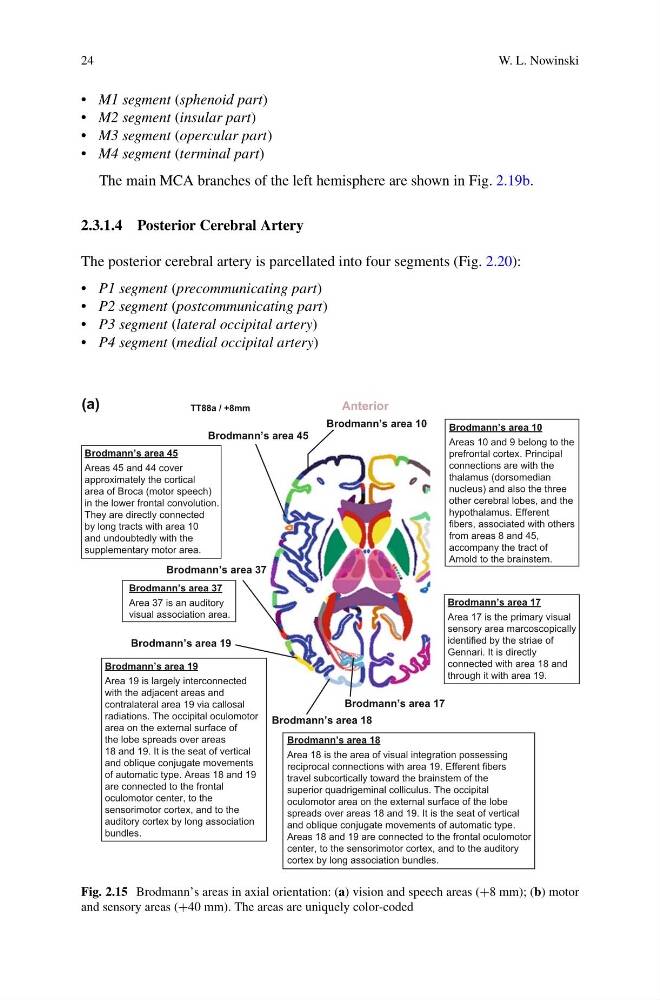
Fig. 2.15 Brodmann’s areas in axial orientation: (a) vision and speech areas (+8 mm); (b) motor
and sensory areas (+40 mm). The areas are uniquely color-coded
W. L. Nowinski
• M1 segment (sphenoid part)
• M2 segment (insular part)
• M3 segment (opercular part)
• M4 segment (terminal part)
The main MCA branches of the left hemisphere are shown in Fig. 2.19b.
2.3.1.4 Posterior Cerebral Artery
The posterior cerebral artery is parcellated into four segments (Fig. 2.20):
• P1 segment (precommunicating part)
• P2 segment (postcommunicating part)
• P3 segment (lateral occipital artery)
• P4 segment (medial occipital artery)
Brodmann’s area 10
Areas 10 and 9 belong to the
prefrontal cortex. Principal
connections are with the
thalamus (dorsomedian
nucleus) and also the three
other cerebral lobes, and the
hypothalamus. Efferent
fibers, associated with others
from areas 8 and 45,
accompany the tract of
Arnold to the brainstem.
Brodmann’s area 10
TT88a / +8mm
(a)
Anterior
Brodmann’s area 45
Areas 45 and 44 cover
approximately the cortical
area of Broca (motor speech)
in the lower frontal convolution.
They are directly connected
by long tracts with area 10
and undoubtedly with the
supplementary motor area.
Brodmann’s area 45
Brodmann’s area 37
Area 37 is an auditory
visual association area.
Brodmann’s area 37
Brodmann’s area 19
Area 19 is largely interconnected
with the adjacent areas and
contralateral area 19 via callosal
radiations. The occipital oculomotor
area on the external surface of
the lobe spreads over areas
18 and 19. It is the seat of vertical
and oblique conjugate movements
of automatic type. Areas 18 and 19
are connected to the frontal
oculomotor center, to the
sensorimotor cortex, and to the
auditory cortex by long association
bundles.
Brodmann’s area 19
Brodmann’s area 18
Area 18 is the area of visual integration possessing
reciprocal connections with area 19. Efferent fibers
travel subcortically toward the brainstem of the
superior quadrigeminal colliculus. The occipital
oculomotor area on the external surface of the lobe
spreads over areas 18 and 19. It is the seat of vertical
and oblique conjugate movements of automatic type.
Areas 18 and 19 are connected to the frontal oculomotor
center, to the sensorimotor cortex, and to the auditory
cortex by long association bundles.
Brodmann’s area 18
Brodmann’s area 17
Brodmann’s area 17
Area 17 is the primary visual
sensory area marcoscopically
identified by the striae of
Gennari. It is directly
connected with area 18 and
through it with area 19.
Fig. 2.15 Brodmann’s areas in axial orientation: (a) vision and speech areas (+8 mm); (b) motor
and sensory areas (+40 mm). The areas are uniquely color-coded