

312
S. Frisken et al.
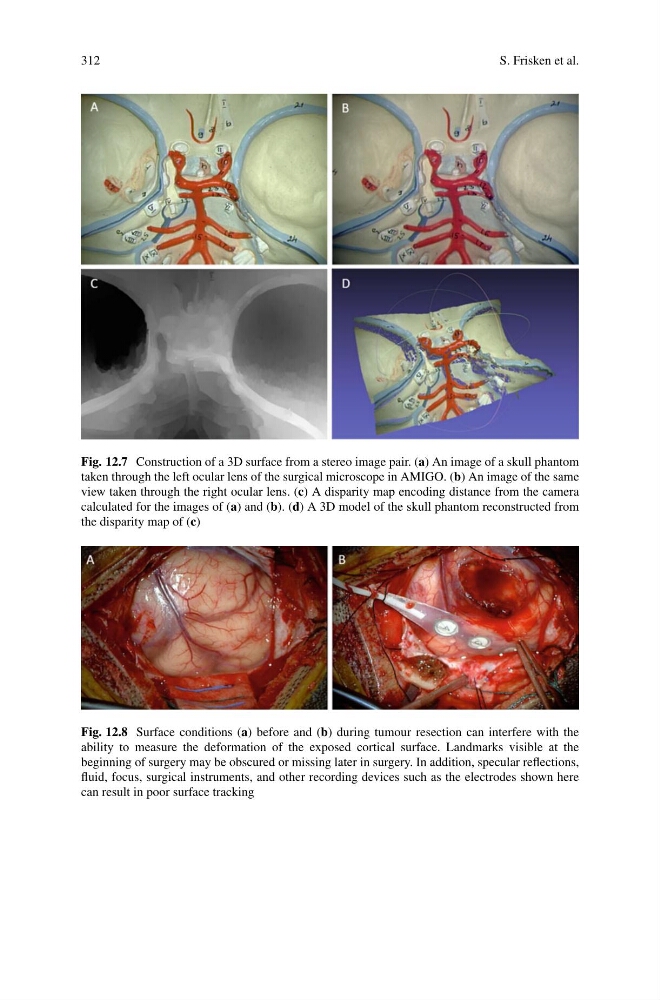
Fig. 12.7 Construction of a 3D surface from a stereo image pair. (a) An image of a skull phantom
taken through the left ocular lens of the surgical microscope in AMIGO. (b) An image of the same
view taken through the right ocular lens. (c) A disparity map encoding distance from the camera
calculated for the images of (a) and (b). (d) A 3D model of the skull phantom reconstructed from
the disparity map of (c)
Fig. 12.8 Surface conditions (a) before and (b) during tumour resection can interfere with the
ability to measure the deformation of the exposed cortical surface. Landmarks visible at the
beginning of surgery may be obscured or missing later in surgery. In addition, specular reflections,
fluid, focus, surgical instruments, and other recording devices such as the electrodes shown here
can result in poor surface tracking
S. Frisken et al.
Fig. 12.7 Construction of a 3D surface from a stereo image pair. (a) An image of a skull phantom
taken through the left ocular lens of the surgical microscope in AMIGO. (b) An image of the same
view taken through the right ocular lens. (c) A disparity map encoding distance from the camera
calculated for the images of (a) and (b). (d) A 3D model of the skull phantom reconstructed from
the disparity map of (c)
Fig. 12.8 Surface conditions (a) before and (b) during tumour resection can interfere with the
ability to measure the deformation of the exposed cortical surface. Landmarks visible at the
beginning of surgery may be obscured or missing later in surgery. In addition, specular reflections,
fluid, focus, surgical instruments, and other recording devices such as the electrodes shown here
can result in poor surface tracking