

308
S. Frisken et al.
iMRI has the advantage that it can provide high-resolution images of the full
brain that are familiar and relatively easy to interpret by neurosurgeons. In addition,
some iMRI systems can provide additional fMRI or DTI. Unfortunately, iMRI
systems are expensive; they require specialised equipment and a specially trained
surgical team. The early vision of performing neurosurgery for tumour resection
in the magnet has been mostly replaced by convenient access to an MRI scanner,
either by transferring the patient or moving the scanner (see Fig. 12.5). While it has
proven effective for improving the extent of resection [41–43], this approach is time
consuming and disruptive to surgery, making it impractical to use iMRI to update
neuronavigation frequently during surgery.
In contrast, intra-operative ultrasound (iUS) only provides a limited field of view
through the craniotomy, is hard to interpret, and is unfamiliar to most neurosurgeons.
On the other hand, it is much less expensive and more readily available than iMRI
and is less disruptive to surgery. A number of groups have used 2D or 3D iUS
for direct navigation during neurosurgery [44–50]. SINTEF and the Norwegian
National Advisory Unit for Ultrasound and Image-Guided Therapy have offered
a course in the use of iUS in neurosurgery since 2008 [51]. This course covers
both direct navigation from iUS and measurement of brain shift using iUS. Steno
et al. [49] recently reported a retrospective study comparing outcomes when using
conventional neuronavigation based on pre-operative MRI that doesn’t account for
brain shift to outcomes when using neuronavigation based on iUS. They found that
the iUS system improved the extent of resection (EOR) with no adverse effects on
outcome. With the iUS system, the median EOR improved from 75.9% to 87.1%,
and the mean EOR improved from 86.8% to 93.5%.
12.2.3.2 Brain Shift Compensation
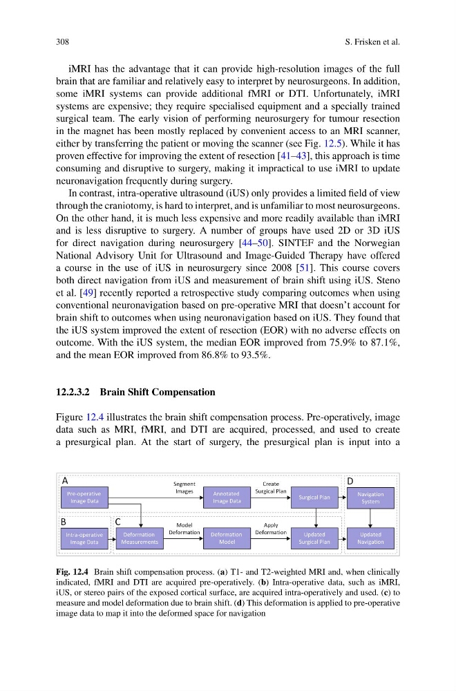
Figure 12.4 illustrates the brain shift compensation process. Pre-operatively, image
data such as MRI, fMRI, and DTI are acquired, processed, and used to create
a presurgical plan. At the start of surgery, the presurgical plan is input into a
Pre-operative
Image Data
Intra-operative
Image Data
Segment
Images
Annotated
Image Data
Deformation
Measurements
Deformation
Model
Create
Surgical Plan
Updated
Surgical Plan
Surgical Plan
A
B
C
Model
Deformation
Updated
Navigation
D
Apply
Deformation
Navigation
System
Fig. 12.4 Brain shift compensation process. (a) T1- and T2-weighted MRI and, when clinically
indicated, fMRI and DTI are acquired pre-operatively. (b) Intra-operative data, such as iMRI,
iUS, or stereo pairs of the exposed cortical surface, are acquired intra-operatively and used. (c) to
measure and model deformation due to brain shift. (d) This deformation is applied to pre-operative
image data to map it into the deformed space for navigation
S. Frisken et al.
iMRI has the advantage that it can provide high-resolution images of the full
brain that are familiar and relatively easy to interpret by neurosurgeons. In addition,
some iMRI systems can provide additional fMRI or DTI. Unfortunately, iMRI
systems are expensive; they require specialised equipment and a specially trained
surgical team. The early vision of performing neurosurgery for tumour resection
in the magnet has been mostly replaced by convenient access to an MRI scanner,
either by transferring the patient or moving the scanner (see Fig. 12.5). While it has
proven effective for improving the extent of resection [41–43], this approach is time
consuming and disruptive to surgery, making it impractical to use iMRI to update
neuronavigation frequently during surgery.
In contrast, intra-operative ultrasound (iUS) only provides a limited field of view
through the craniotomy, is hard to interpret, and is unfamiliar to most neurosurgeons.
On the other hand, it is much less expensive and more readily available than iMRI
and is less disruptive to surgery. A number of groups have used 2D or 3D iUS
for direct navigation during neurosurgery [44–50]. SINTEF and the Norwegian
National Advisory Unit for Ultrasound and Image-Guided Therapy have offered
a course in the use of iUS in neurosurgery since 2008 [51]. This course covers
both direct navigation from iUS and measurement of brain shift using iUS. Steno
et al. [49] recently reported a retrospective study comparing outcomes when using
conventional neuronavigation based on pre-operative MRI that doesn’t account for
brain shift to outcomes when using neuronavigation based on iUS. They found that
the iUS system improved the extent of resection (EOR) with no adverse effects on
outcome. With the iUS system, the median EOR improved from 75.9% to 87.1%,
and the mean EOR improved from 86.8% to 93.5%.
12.2.3.2 Brain Shift Compensation
Figure 12.4 illustrates the brain shift compensation process. Pre-operatively, image
data such as MRI, fMRI, and DTI are acquired, processed, and used to create
a presurgical plan. At the start of surgery, the presurgical plan is input into a
Pre-operative
Image Data
Intra-operative
Image Data
Segment
Images
Annotated
Image Data
Deformation
Measurements
Deformation
Model
Create
Surgical Plan
Updated
Surgical Plan
Surgical Plan
A
B
C
Model
Deformation
Updated
Navigation
D
Apply
Deformation
Navigation
System
Fig. 12.4 Brain shift compensation process. (a) T1- and T2-weighted MRI and, when clinically
indicated, fMRI and DTI are acquired pre-operatively. (b) Intra-operative data, such as iMRI,
iUS, or stereo pairs of the exposed cortical surface, are acquired intra-operatively and used. (c) to
measure and model deformation due to brain shift. (d) This deformation is applied to pre-operative
image data to map it into the deformed space for navigation