

12 Intra-operative Measurement of Brain Deformation
307
12.2.3 Neuronavigation with Brain Shift
There are two basic approaches for updating neuronavigation during surgery in
the presence of brain shift. The first approach replaces pre-operative images with
images acquired during surgery and allows neurosurgeons to navigate from these
new images. The second approach, known as brain shift compensation, measures
brain shift intra-operatively and uses these measurements to update pre-operative
images (e.g. by deforming them to match the brain-shifted brain).
12.2.3.1 Replacing Pre-operative Images with Intra-operative Images
In the late 1990s and early 2000s, a number of intra-operative MRI (iMRI) systems
were developed which combined MRI imaging with surgical access to the patient.
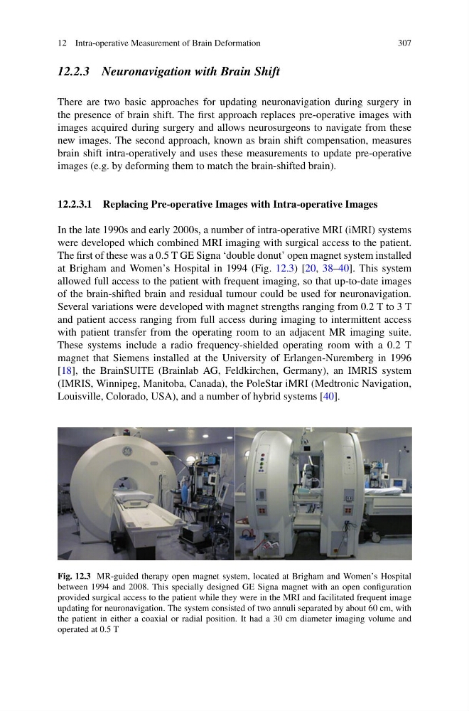
The first of these was a 0.5 T GE Signa ‘double donut’ open magnet system installed
at Brigham and Women’s Hospital in 1994 (Fig. 12.3) [20, 38–40]. This system
allowed full access to the patient with frequent imaging, so that up-to-date images
of the brain-shifted brain and residual tumour could be used for neuronavigation.
Several variations were developed with magnet strengths ranging from 0.2 T to 3 T
and patient access ranging from full access during imaging to intermittent access
with patient transfer from the operating room to an adjacent MR imaging suite.
These systems include a radio frequency-shielded operating room with a 0.2 T
magnet that Siemens installed at the University of Erlangen-Nuremberg in 1996
[18], the BrainSUITE (Brainlab AG, Feldkirchen, Germany), an IMRIS system
(IMRIS, Winnipeg, Manitoba, Canada), the PoleStar iMRI (Medtronic Navigation,
Louisville, Colorado, USA), and a number of hybrid systems [40].
Fig. 12.3 MR-guided therapy open magnet system, located at Brigham and Women’s Hospital
between 1994 and 2008. This specially designed GE Signa magnet with an open configuration
provided surgical access to the patient while they were in the MRI and facilitated frequent image
updating for neuronavigation. The system consisted of two annuli separated by about 60 cm, with
the patient in either a coaxial or radial position. It had a 30 cm diameter imaging volume and
operated at 0.5 T
307
12.2.3 Neuronavigation with Brain Shift
There are two basic approaches for updating neuronavigation during surgery in
the presence of brain shift. The first approach replaces pre-operative images with
images acquired during surgery and allows neurosurgeons to navigate from these
new images. The second approach, known as brain shift compensation, measures
brain shift intra-operatively and uses these measurements to update pre-operative
images (e.g. by deforming them to match the brain-shifted brain).
12.2.3.1 Replacing Pre-operative Images with Intra-operative Images
In the late 1990s and early 2000s, a number of intra-operative MRI (iMRI) systems
were developed which combined MRI imaging with surgical access to the patient.
The first of these was a 0.5 T GE Signa ‘double donut’ open magnet system installed
at Brigham and Women’s Hospital in 1994 (Fig. 12.3) [20, 38–40]. This system
allowed full access to the patient with frequent imaging, so that up-to-date images
of the brain-shifted brain and residual tumour could be used for neuronavigation.
Several variations were developed with magnet strengths ranging from 0.2 T to 3 T
and patient access ranging from full access during imaging to intermittent access
with patient transfer from the operating room to an adjacent MR imaging suite.
These systems include a radio frequency-shielded operating room with a 0.2 T
magnet that Siemens installed at the University of Erlangen-Nuremberg in 1996
[18], the BrainSUITE (Brainlab AG, Feldkirchen, Germany), an IMRIS system
(IMRIS, Winnipeg, Manitoba, Canada), the PoleStar iMRI (Medtronic Navigation,
Louisville, Colorado, USA), and a number of hybrid systems [40].
Fig. 12.3 MR-guided therapy open magnet system, located at Brigham and Women’s Hospital
between 1994 and 2008. This specially designed GE Signa magnet with an open configuration
provided surgical access to the patient while they were in the MRI and facilitated frequent image
updating for neuronavigation. The system consisted of two annuli separated by about 60 cm, with
the patient in either a coaxial or radial position. It had a 30 cm diameter imaging volume and
operated at 0.5 T