

12 Intra-operative Measurement of Brain Deformation
305
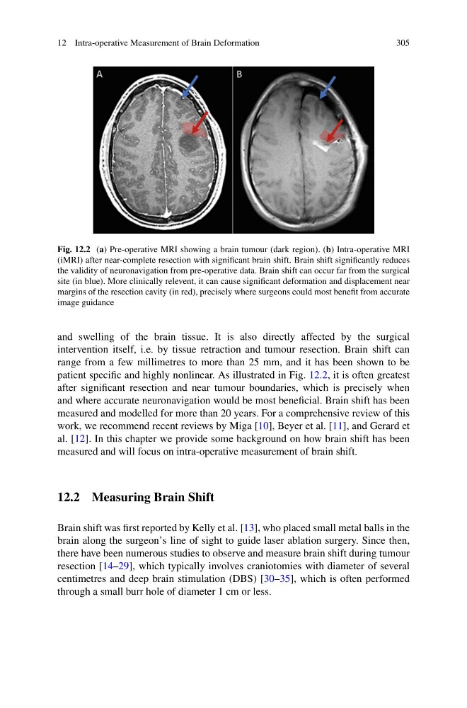
Fig. 12.2 (a) Pre-operative MRI showing a brain tumour (dark region). (b) Intra-operative MRI
(iMRI) after near-complete resection with significant brain shift. Brain shift significantly reduces
the validity of neuronavigation from pre-operative data. Brain shift can occur far from the surgical
site (in blue). More clinically relevent, it can cause significant deformation and displacement near
margins of the resection cavity (in red), precisely where surgeons could most benefit from accurate
image guidance
and swelling of the brain tissue. It is also directly affected by the surgical
intervention itself, i.e. by tissue retraction and tumour resection. Brain shift can
range from a few millimetres to more than 25 mm, and it has been shown to be
patient specific and highly nonlinear. As illustrated in Fig. 12.2, it is often greatest
after significant resection and near tumour boundaries, which is precisely when
and where accurate neuronavigation would be most beneficial. Brain shift has been
measured and modelled for more than 20 years. For a comprehensive review of this
work, we recommend recent reviews by Miga [10], Beyer et al. [11], and Gerard et
al. [12]. In this chapter we provide some background on how brain shift has been
measured and will focus on intra-operative measurement of brain shift.
12.2 Measuring Brain Shift
Brain shift was first reported by Kelly et al. [13], who placed small metal balls in the
brain along the surgeon’s line of sight to guide laser ablation surgery. Since then,
there have been numerous studies to observe and measure brain shift during tumour
resection [14–29], which typically involves craniotomies with diameter of several
centimetres and deep brain stimulation (DBS) [30–35], which is often performed
through a small burr hole of diameter 1 cm or less.
305
Fig. 12.2 (a) Pre-operative MRI showing a brain tumour (dark region). (b) Intra-operative MRI
(iMRI) after near-complete resection with significant brain shift. Brain shift significantly reduces
the validity of neuronavigation from pre-operative data. Brain shift can occur far from the surgical
site (in blue). More clinically relevent, it can cause significant deformation and displacement near
margins of the resection cavity (in red), precisely where surgeons could most benefit from accurate
image guidance
and swelling of the brain tissue. It is also directly affected by the surgical
intervention itself, i.e. by tissue retraction and tumour resection. Brain shift can
range from a few millimetres to more than 25 mm, and it has been shown to be
patient specific and highly nonlinear. As illustrated in Fig. 12.2, it is often greatest
after significant resection and near tumour boundaries, which is precisely when
and where accurate neuronavigation would be most beneficial. Brain shift has been
measured and modelled for more than 20 years. For a comprehensive review of this
work, we recommend recent reviews by Miga [10], Beyer et al. [11], and Gerard et
al. [12]. In this chapter we provide some background on how brain shift has been
measured and will focus on intra-operative measurement of brain shift.
12.2 Measuring Brain Shift
Brain shift was first reported by Kelly et al. [13], who placed small metal balls in the
brain along the surgeon’s line of sight to guide laser ablation surgery. Since then,
there have been numerous studies to observe and measure brain shift during tumour
resection [14–29], which typically involves craniotomies with diameter of several
centimetres and deep brain stimulation (DBS) [30–35], which is often performed
through a small burr hole of diameter 1 cm or less.