

304
S. Frisken et al.
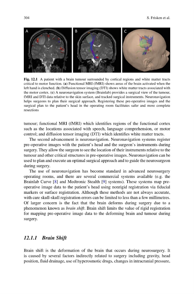
Fig. 12.1 A patient with a brain tumour surrounded by cortical regions and white matter tracts
critical to motor function. (a) Functional MRI (fMRI) shows areas of the brain activated when the
left hand is clenched. (b) Diffusion tensor imaging (DTI) shows white matter tracts associated with
the motor cortex. (c) A neuronavigation system (Brainlab) provides a surgical view of the tumour,
fMRI and DTI data relative to the skin surface, and tracked surgical instruments. Neuronavigation
helps surgeons to plan their surgical approach. Registering these pre-operative images and the
surgical plan to the patient’s head in the operating room facilitates safer and more complete
resections
tumour; functional MRI (fMRI) which identifies regions of the functional cortex
such as the locations associated with speech, language comprehension, or motor
control; and diffusion tensor imaging (DTI) which identifies white matter tracts.
The second advancement is neuronavigation. Neuronavigation systems register
pre-operative images with the patient’s head and the surgeon’s instruments during
surgery. They allow the surgeon to see the location of their instruments relative to the
tumour and other critical structures in pre-operative images. Neuronavigation can be
used to plan and execute an optimal surgical approach and to guide the neurosurgeon
during surgery.
The use of neuronavigation has become standard in advanced neurosurgery
operating rooms, and there are several commercial systems available (e.g. the
Brainlab Curve [8] and Medtronic Stealth [9] systems). These systems map preoperative image data to the patient’s head using nonrigid registration via fiducial
markers or surface registration. Although these methods are not always accurate,
with care skull-skull registration errors can be limited to less than a few millimetres.
Of larger concern is the fact that the brain deforms during surgery due to a
phenomenon known as brain shift. Brain shift limits the value of rigid registration
for mapping pre-operative image data to the deforming brain and tumour during
surgery.
12.1.1 Brain Shift
Brain shift is the deformation of the brain that occurs during neurosurgery. It
is caused by several factors indirectly related to surgery including gravity, head
position, fluid drainage, use of hyperosmotic drugs, changes in intracranial pressure,
S. Frisken et al.
Fig. 12.1 A patient with a brain tumour surrounded by cortical regions and white matter tracts
critical to motor function. (a) Functional MRI (fMRI) shows areas of the brain activated when the
left hand is clenched. (b) Diffusion tensor imaging (DTI) shows white matter tracts associated with
the motor cortex. (c) A neuronavigation system (Brainlab) provides a surgical view of the tumour,
fMRI and DTI data relative to the skin surface, and tracked surgical instruments. Neuronavigation
helps surgeons to plan their surgical approach. Registering these pre-operative images and the
surgical plan to the patient’s head in the operating room facilitates safer and more complete
resections
tumour; functional MRI (fMRI) which identifies regions of the functional cortex
such as the locations associated with speech, language comprehension, or motor
control; and diffusion tensor imaging (DTI) which identifies white matter tracts.
The second advancement is neuronavigation. Neuronavigation systems register
pre-operative images with the patient’s head and the surgeon’s instruments during
surgery. They allow the surgeon to see the location of their instruments relative to the
tumour and other critical structures in pre-operative images. Neuronavigation can be
used to plan and execute an optimal surgical approach and to guide the neurosurgeon
during surgery.
The use of neuronavigation has become standard in advanced neurosurgery
operating rooms, and there are several commercial systems available (e.g. the
Brainlab Curve [8] and Medtronic Stealth [9] systems). These systems map preoperative image data to the patient’s head using nonrigid registration via fiducial
markers or surface registration. Although these methods are not always accurate,
with care skull-skull registration errors can be limited to less than a few millimetres.
Of larger concern is the fact that the brain deforms during surgery due to a
phenomenon known as brain shift. Brain shift limits the value of rigid registration
for mapping pre-operative image data to the deforming brain and tumour during
surgery.
12.1.1 Brain Shift
Brain shift is the deformation of the brain that occurs during neurosurgery. It
is caused by several factors indirectly related to surgery including gravity, head
position, fluid drainage, use of hyperosmotic drugs, changes in intracranial pressure,