

220
V. Kurtcuoglu et al.
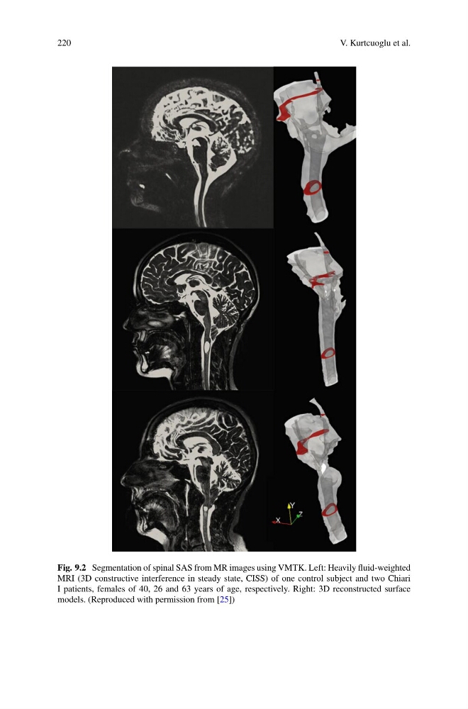
Fig. 9.2 Segmentation of spinal SAS from MR images using VMTK. Left: Heavily fluid-weighted
MRI (3D constructive interference in steady state, CISS) of one control subject and two Chiari
I patients, females of 40, 26 and 63 years of age, respectively. Right: 3D reconstructed surface
models. (Reproduced with permission from [25])
V. Kurtcuoglu et al.
Fig. 9.2 Segmentation of spinal SAS from MR images using VMTK. Left: Heavily fluid-weighted
MRI (3D constructive interference in steady state, CISS) of one control subject and two Chiari
I patients, females of 40, 26 and 63 years of age, respectively. Right: 3D reconstructed surface
models. (Reproduced with permission from [25])