

8 Dynamics of Cerebrospinal Fluid: From Theoretical Models to Clinical Applications
201
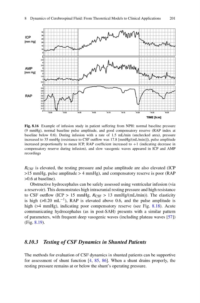
Fig. 8.16 Example of infusion study in patient suffering from NPH: normal baseline pressure
(9 mmHg), normal baseline pulse amplitude, and good compensatory reserve (RAP index at
baseline below 0.6). During infusion with a rate of 1.5 mL/min (unchecked area), pressure
increased to 35 mmHg (resistance to CSF outflow was 17.8 [mmHg/(mL/min)]), pulse amplitude
increased proportionally to mean ICP, RAP coefficient increased to +1 (indicating decrease in
compensatory reserve during infusion), and slow vasogenic waves appeared in ICP and AMP
recordings
R CSF is elevated, the resting pressure and pulse amplitude are also elevated (ICP
>15 mmHg, pulse amplitude > 4 mmHg), and compensatory reserve is poor (RAP
>0.6 at baseline).
Obstructive hydrocephalus can be safely assessed using ventricular infusion (via
a reservoir). This demonstrates high intracranial resting pressure and high resistance
to CSF outflow (ICP > 15 mmHg, R CSF > 13 mmHg/(mL/min)). The elasticity
is high (>0.20 mL −1 ), RAP is elevated above 0.6, and the pulse amplitude is
high (>4 mmHg), indicating poor compensatory reserve (see Fig. 8.18). Acute
communicating hydrocephalus (as in post-SAH) presents with a similar pattern
of parameters, with frequent deep vasogenic waves (including plateau waves [57])
(Fig. 8.19).
8.10.3 Testing of CSF Dynamics in Shunted Patients
The methods for evaluation of CSF dynamics in shunted patients can be supportive
for assessment of shunt function [4, 85, 86]. When a shunt drains properly, the
resting pressure remains at or below the shunt’s operating pressure.
201
Fig. 8.16 Example of infusion study in patient suffering from NPH: normal baseline pressure
(9 mmHg), normal baseline pulse amplitude, and good compensatory reserve (RAP index at
baseline below 0.6). During infusion with a rate of 1.5 mL/min (unchecked area), pressure
increased to 35 mmHg (resistance to CSF outflow was 17.8 [mmHg/(mL/min)]), pulse amplitude
increased proportionally to mean ICP, RAP coefficient increased to +1 (indicating decrease in
compensatory reserve during infusion), and slow vasogenic waves appeared in ICP and AMP
recordings
R CSF is elevated, the resting pressure and pulse amplitude are also elevated (ICP
>15 mmHg, pulse amplitude > 4 mmHg), and compensatory reserve is poor (RAP
>0.6 at baseline).
Obstructive hydrocephalus can be safely assessed using ventricular infusion (via
a reservoir). This demonstrates high intracranial resting pressure and high resistance
to CSF outflow (ICP > 15 mmHg, R CSF > 13 mmHg/(mL/min)). The elasticity
is high (>0.20 mL −1 ), RAP is elevated above 0.6, and the pulse amplitude is
high (>4 mmHg), indicating poor compensatory reserve (see Fig. 8.18). Acute
communicating hydrocephalus (as in post-SAH) presents with a similar pattern
of parameters, with frequent deep vasogenic waves (including plateau waves [57])
(Fig. 8.19).
8.10.3 Testing of CSF Dynamics in Shunted Patients
The methods for evaluation of CSF dynamics in shunted patients can be supportive
for assessment of shunt function [4, 85, 86]. When a shunt drains properly, the
resting pressure remains at or below the shunt’s operating pressure.