

8 Dynamics of Cerebrospinal Fluid: From Theoretical Models to Clinical Applications
199
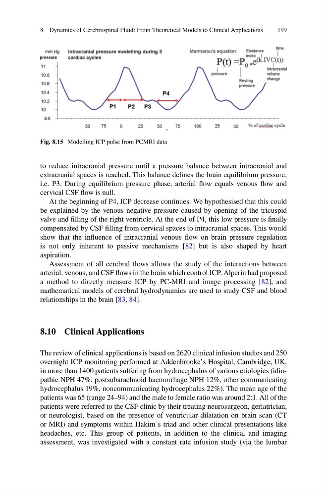
Fig. 8.15 Modelling ICP pulse from PCMRI data
to reduce intracranial pressure until a pressure balance between intracranial and
extracranial spaces is reached. This balance defines the brain equilibrium pressure,
i.e. P3. During equilibrium pressure phase, arterial flow equals venous flow and
cervical CSF flow is null.
At the beginning of P4, ICP decrease continues. We hypothesised that this could
be explained by the venous negative pressure caused by opening of the tricuspid
valve and filling of the right ventricle. At the end of P4, this low pressure is finally
compensated by CSF filling from cervical spaces to intracranial spaces. This would
show that the influence of intracranial venous flow on brain pressure regulation
is not only inherent to passive mechanisms [82] but is also shaped by heart
aspiration.
Assessment of all cerebral flows allows the study of the interactions between
arterial, venous, and CSF flows in the brain which control ICP. Alperin had proposed
a method to directly measure ICP by PC-MRI and image processing [82], and
mathematical models of cerebral hydrodynamics are used to study CSF and blood
relationships in the brain [83, 84].
8.10 Clinical Applications
The review of clinical applications is based on 2620 clinical infusion studies and 250
overnight ICP monitoring performed at Addenbrooke’s Hospital, Cambridge, UK,
in more than 1400 patients suffering from hydrocephalus of various etiologies (idiopathic NPH 47%, postsubarachnoid haemorrhage NPH 12%, other communicating
hydrocephalus 19%, noncommunicating hydrocephalus 22%). The mean age of the
patients was 65 (range 24–94) and the male to female ratio was around 2:1. All of the
patients were referred to the CSF clinic by their treating neurosurgeon, geriatrician,
or neurologist, based on the presence of ventricular dilatation on brain scan (CT
or MRI) and symptoms within Hakim’s triad and other clinical presentations like
headaches, etc. This group of patients, in addition to the clinical and imaging
assessment, was investigated with a constant rate infusion study (via the lumbar
199
Fig. 8.15 Modelling ICP pulse from PCMRI data
to reduce intracranial pressure until a pressure balance between intracranial and
extracranial spaces is reached. This balance defines the brain equilibrium pressure,
i.e. P3. During equilibrium pressure phase, arterial flow equals venous flow and
cervical CSF flow is null.
At the beginning of P4, ICP decrease continues. We hypothesised that this could
be explained by the venous negative pressure caused by opening of the tricuspid
valve and filling of the right ventricle. At the end of P4, this low pressure is finally
compensated by CSF filling from cervical spaces to intracranial spaces. This would
show that the influence of intracranial venous flow on brain pressure regulation
is not only inherent to passive mechanisms [82] but is also shaped by heart
aspiration.
Assessment of all cerebral flows allows the study of the interactions between
arterial, venous, and CSF flows in the brain which control ICP. Alperin had proposed
a method to directly measure ICP by PC-MRI and image processing [82], and
mathematical models of cerebral hydrodynamics are used to study CSF and blood
relationships in the brain [83, 84].
8.10 Clinical Applications
The review of clinical applications is based on 2620 clinical infusion studies and 250
overnight ICP monitoring performed at Addenbrooke’s Hospital, Cambridge, UK,
in more than 1400 patients suffering from hydrocephalus of various etiologies (idiopathic NPH 47%, postsubarachnoid haemorrhage NPH 12%, other communicating
hydrocephalus 19%, noncommunicating hydrocephalus 22%). The mean age of the
patients was 65 (range 24–94) and the male to female ratio was around 2:1. All of the
patients were referred to the CSF clinic by their treating neurosurgeon, geriatrician,
or neurologist, based on the presence of ventricular dilatation on brain scan (CT
or MRI) and symptoms within Hakim’s triad and other clinical presentations like
headaches, etc. This group of patients, in addition to the clinical and imaging
assessment, was investigated with a constant rate infusion study (via the lumbar