

7 Biomechanical Modelling of the Brain for Neuronavigation in Epilepsy Surgery
169
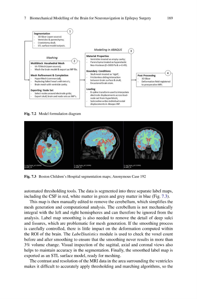
Fig. 7.2 Model formulation diagram
Fig. 7.3 Boston Children’s Hospital segmentation maps; Anonymous Case 192
automated thresholding tools. The data is segmented into three separate label maps,
including the CSF in red, white matter in green and grey matter in blue (Fig. 7.3).
This map is then manually edited to remove the cerebellum, which simplifies the
mesh generation and computational analysis. The cerebellum is not mechanically
integral with the left and right hemispheres and can therefore be ignored from the
analysis. Label map smoothing is also needed to remove the detail of deep sulci
and fissures, which are problematic for mesh generation. If the smoothing process
is carefully controlled, there is little impact on the deformation computed within
the ROI of the brain. The LabelStatistics module is used to check the voxel count
before and after smoothing to ensure that the smoothing never results in more than
3% volume change. Visual inspection of the sagittal, axial and coronal views also
helps to maintain accuracy in the segmentation. Finally, the smoothed label map is
exported as an STL surface model, ready for meshing.
The contrast and resolution of the MRI data in the area surrounding the ventricles
makes it difficult to accurately apply thresholding and marching algorithms, so the
169
Fig. 7.2 Model formulation diagram
Fig. 7.3 Boston Children’s Hospital segmentation maps; Anonymous Case 192
automated thresholding tools. The data is segmented into three separate label maps,
including the CSF in red, white matter in green and grey matter in blue (Fig. 7.3).
This map is then manually edited to remove the cerebellum, which simplifies the
mesh generation and computational analysis. The cerebellum is not mechanically
integral with the left and right hemispheres and can therefore be ignored from the
analysis. Label map smoothing is also needed to remove the detail of deep sulci
and fissures, which are problematic for mesh generation. If the smoothing process
is carefully controlled, there is little impact on the deformation computed within
the ROI of the brain. The LabelStatistics module is used to check the voxel count
before and after smoothing to ensure that the smoothing never results in more than
3% volume change. Visual inspection of the sagittal, axial and coronal views also
helps to maintain accuracy in the segmentation. Finally, the smoothed label map is
exported as an STL surface model, ready for meshing.
The contrast and resolution of the MRI data in the area surrounding the ventricles
makes it difficult to accurately apply thresholding and marching algorithms, so the