

138
K. Miller et al.
specific biomechanical models quickly and reliably remains unresolved (see [1]
and Chap. 7 for current attempts to address this issue using meshless methods).
Another aspect worth considering is that for computational biomechanics to be
accepted and beneficial in clinical practice, biomechanical computations must be
seamlessly incorporated into the clinical work flow. This can only be achieved if
these computations are conducted at least in close to real time (how to achieve this
is discussed in Chap. 2; see also [11]).
In the remainder of this section, we will discuss the main modelling issues:
geometry, boundary conditions, loading and tissue mechanical properties.
6.2.1 Geometry
Detailed geometric information is needed to define the domain in which the
deformation field needs to be computed. Such information is provided by electronic
brain atlases described in detail in Chap. 2 and is readily available as some atlases
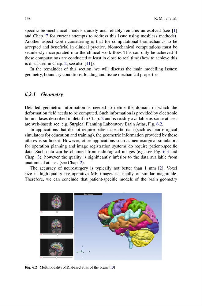
are web-based; see, e.g. Surgical Planning Laboratory Brain Atlas, Fig. 6.2.
In applications that do not require patient-specific data (such as neurosurgical
simulators for education and training), the geometric information provided by these
atlases is sufficient. However, other applications such as neurosurgical simulators
for operation planning and image registration systems do require patient-specific
data. Such data can be obtained from radiological images (e.g. see Fig. 6.3 and
Chap. 3); however the quality is significantly inferior to the data available from
anatomical atlases (see Chap. 2).
The accuracy of neurosurgery is typically not better than 1 mm [2]. Voxel
size in high-quality pre-operative MR images is usually of similar magnitude.
Therefore, we can conclude that patient-specific models of the brain geometry
Fig. 6.2 Multimodality MRI-based atlas of the brain [13]
K. Miller et al.
specific biomechanical models quickly and reliably remains unresolved (see [1]
and Chap. 7 for current attempts to address this issue using meshless methods).
Another aspect worth considering is that for computational biomechanics to be
accepted and beneficial in clinical practice, biomechanical computations must be
seamlessly incorporated into the clinical work flow. This can only be achieved if
these computations are conducted at least in close to real time (how to achieve this
is discussed in Chap. 2; see also [11]).
In the remainder of this section, we will discuss the main modelling issues:
geometry, boundary conditions, loading and tissue mechanical properties.
6.2.1 Geometry
Detailed geometric information is needed to define the domain in which the
deformation field needs to be computed. Such information is provided by electronic
brain atlases described in detail in Chap. 2 and is readily available as some atlases
are web-based; see, e.g. Surgical Planning Laboratory Brain Atlas, Fig. 6.2.
In applications that do not require patient-specific data (such as neurosurgical
simulators for education and training), the geometric information provided by these
atlases is sufficient. However, other applications such as neurosurgical simulators
for operation planning and image registration systems do require patient-specific
data. Such data can be obtained from radiological images (e.g. see Fig. 6.3 and
Chap. 3); however the quality is significantly inferior to the data available from
anatomical atlases (see Chap. 2).
The accuracy of neurosurgery is typically not better than 1 mm [2]. Voxel
size in high-quality pre-operative MR images is usually of similar magnitude.
Therefore, we can conclude that patient-specific models of the brain geometry
Fig. 6.2 Multimodality MRI-based atlas of the brain [13]