

120
K. H. Yang and H. Mao
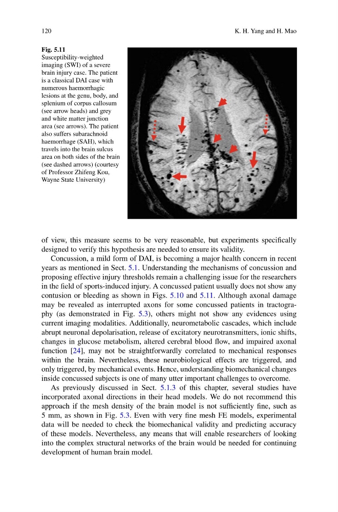
Fig. 5.11
Susceptibility-weighted
imaging (SWI) of a severe
brain injury case. The patient
is a classical DAI case with
numerous haemorrhagic
lesions at the genu, body, and
splenium of corpus callosum
(see arrow heads) and grey
and white matter junction
area (see arrows). The patient
also suffers subarachnoid
haemorrhage (SAH), which
travels into the brain sulcus
area on both sides of the brain
(see dashed arrows) (courtesy
of Professor Zhifeng Kou,
Wayne State University)
of view, this measure seems to be very reasonable, but experiments specifically
designed to verify this hypothesis are needed to ensure its validity.
Concussion, a mild form of DAI, is becoming a major health concern in recent
years as mentioned in Sect. 5.1. Understanding the mechanisms of concussion and
proposing effective injury thresholds remain a challenging issue for the researchers
in the field of sports-induced injury. A concussed patient usually does not show any
contusion or bleeding as shown in Figs. 5.10 and 5.11. Although axonal damage
may be revealed as interrupted axons for some concussed patients in tractography (as demonstrated in Fig. 5.3), others might not show any evidences using
current imaging modalities. Additionally, neurometabolic cascades, which include
abrupt neuronal depolarisation, release of excitatory neurotransmitters, ionic shifts,
changes in glucose metabolism, altered cerebral blood flow, and impaired axonal
function [24], may not be straightforwardly correlated to mechanical responses
within the brain. Nevertheless, these neurobiological effects are triggered, and
only triggered, by mechanical events. Hence, understanding biomechanical changes
inside concussed subjects is one of many utter important challenges to overcome.
As previously discussed in Sect. 5.1.3 of this chapter, several studies have
incorporated axonal directions in their head models. We do not recommend this
approach if the mesh density of the brain model is not sufficiently fine, such as
5 mm, as shown in Fig. 5.3. Even with very fine mesh FE models, experimental
data will be needed to check the biomechanical validity and predicting accuracy
of these models. Nevertheless, any means that will enable researchers of looking
into the complex structural networks of the brain would be needed for continuing
development of human brain model.
K. H. Yang and H. Mao
Fig. 5.11
Susceptibility-weighted
imaging (SWI) of a severe
brain injury case. The patient
is a classical DAI case with
numerous haemorrhagic
lesions at the genu, body, and
splenium of corpus callosum
(see arrow heads) and grey
and white matter junction
area (see arrows). The patient
also suffers subarachnoid
haemorrhage (SAH), which
travels into the brain sulcus
area on both sides of the brain
(see dashed arrows) (courtesy
of Professor Zhifeng Kou,
Wayne State University)
of view, this measure seems to be very reasonable, but experiments specifically
designed to verify this hypothesis are needed to ensure its validity.
Concussion, a mild form of DAI, is becoming a major health concern in recent
years as mentioned in Sect. 5.1. Understanding the mechanisms of concussion and
proposing effective injury thresholds remain a challenging issue for the researchers
in the field of sports-induced injury. A concussed patient usually does not show any
contusion or bleeding as shown in Figs. 5.10 and 5.11. Although axonal damage
may be revealed as interrupted axons for some concussed patients in tractography (as demonstrated in Fig. 5.3), others might not show any evidences using
current imaging modalities. Additionally, neurometabolic cascades, which include
abrupt neuronal depolarisation, release of excitatory neurotransmitters, ionic shifts,
changes in glucose metabolism, altered cerebral blood flow, and impaired axonal
function [24], may not be straightforwardly correlated to mechanical responses
within the brain. Nevertheless, these neurobiological effects are triggered, and
only triggered, by mechanical events. Hence, understanding biomechanical changes
inside concussed subjects is one of many utter important challenges to overcome.
As previously discussed in Sect. 5.1.3 of this chapter, several studies have
incorporated axonal directions in their head models. We do not recommend this
approach if the mesh density of the brain model is not sufficiently fine, such as
5 mm, as shown in Fig. 5.3. Even with very fine mesh FE models, experimental
data will be needed to check the biomechanical validity and predicting accuracy
of these models. Nevertheless, any means that will enable researchers of looking
into the complex structural networks of the brain would be needed for continuing
development of human brain model.