

5 Modelling of the Brain for Injury Simulation and Prevention
119
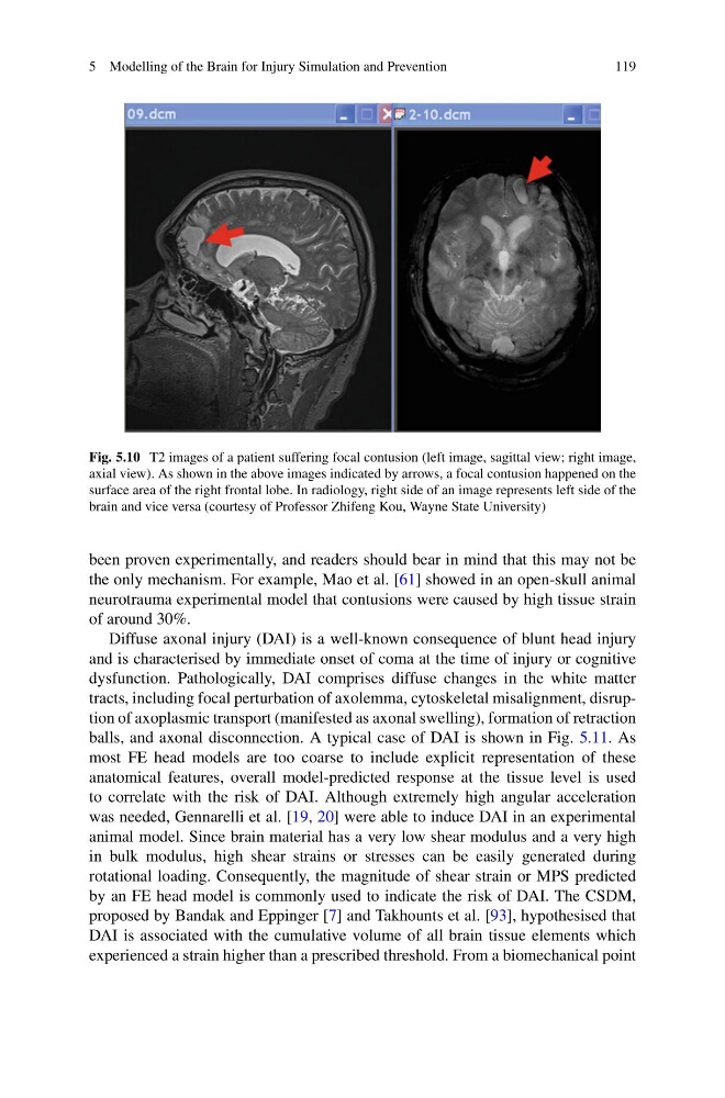
Fig. 5.10 T2 images of a patient suffering focal contusion (left image, sagittal view; right image,
axial view). As shown in the above images indicated by arrows, a focal contusion happened on the
surface area of the right frontal lobe. In radiology, right side of an image represents left side of the
brain and vice versa (courtesy of Professor Zhifeng Kou, Wayne State University)
been proven experimentally, and readers should bear in mind that this may not be
the only mechanism. For example, Mao et al. [61] showed in an open-skull animal
neurotrauma experimental model that contusions were caused by high tissue strain
of around 30%.
Diffuse axonal injury (DAI) is a well-known consequence of blunt head injury
and is characterised by immediate onset of coma at the time of injury or cognitive
dysfunction. Pathologically, DAI comprises diffuse changes in the white matter
tracts, including focal perturbation of axolemma, cytoskeletal misalignment, disruption of axoplasmic transport (manifested as axonal swelling), formation of retraction
balls, and axonal disconnection. A typical case of DAI is shown in Fig. 5.11. As
most FE head models are too coarse to include explicit representation of these
anatomical features, overall model-predicted response at the tissue level is used
to correlate with the risk of DAI. Although extremely high angular acceleration
was needed, Gennarelli et al. [19, 20] were able to induce DAI in an experimental
animal model. Since brain material has a very low shear modulus and a very high
in bulk modulus, high shear strains or stresses can be easily generated during
rotational loading. Consequently, the magnitude of shear strain or MPS predicted
by an FE head model is commonly used to indicate the risk of DAI. The CSDM,
proposed by Bandak and Eppinger [7] and Takhounts et al. [93], hypothesised that
DAI is associated with the cumulative volume of all brain tissue elements which
experienced a strain higher than a prescribed threshold. From a biomechanical point
119
Fig. 5.10 T2 images of a patient suffering focal contusion (left image, sagittal view; right image,
axial view). As shown in the above images indicated by arrows, a focal contusion happened on the
surface area of the right frontal lobe. In radiology, right side of an image represents left side of the
brain and vice versa (courtesy of Professor Zhifeng Kou, Wayne State University)
been proven experimentally, and readers should bear in mind that this may not be
the only mechanism. For example, Mao et al. [61] showed in an open-skull animal
neurotrauma experimental model that contusions were caused by high tissue strain
of around 30%.
Diffuse axonal injury (DAI) is a well-known consequence of blunt head injury
and is characterised by immediate onset of coma at the time of injury or cognitive
dysfunction. Pathologically, DAI comprises diffuse changes in the white matter
tracts, including focal perturbation of axolemma, cytoskeletal misalignment, disruption of axoplasmic transport (manifested as axonal swelling), formation of retraction
balls, and axonal disconnection. A typical case of DAI is shown in Fig. 5.11. As
most FE head models are too coarse to include explicit representation of these
anatomical features, overall model-predicted response at the tissue level is used
to correlate with the risk of DAI. Although extremely high angular acceleration
was needed, Gennarelli et al. [19, 20] were able to induce DAI in an experimental
animal model. Since brain material has a very low shear modulus and a very high
in bulk modulus, high shear strains or stresses can be easily generated during
rotational loading. Consequently, the magnitude of shear strain or MPS predicted
by an FE head model is commonly used to indicate the risk of DAI. The CSDM,
proposed by Bandak and Eppinger [7] and Takhounts et al. [93], hypothesised that
DAI is associated with the cumulative volume of all brain tissue elements which
experienced a strain higher than a prescribed threshold. From a biomechanical point