

5 Modelling of the Brain for Injury Simulation and Prevention
107
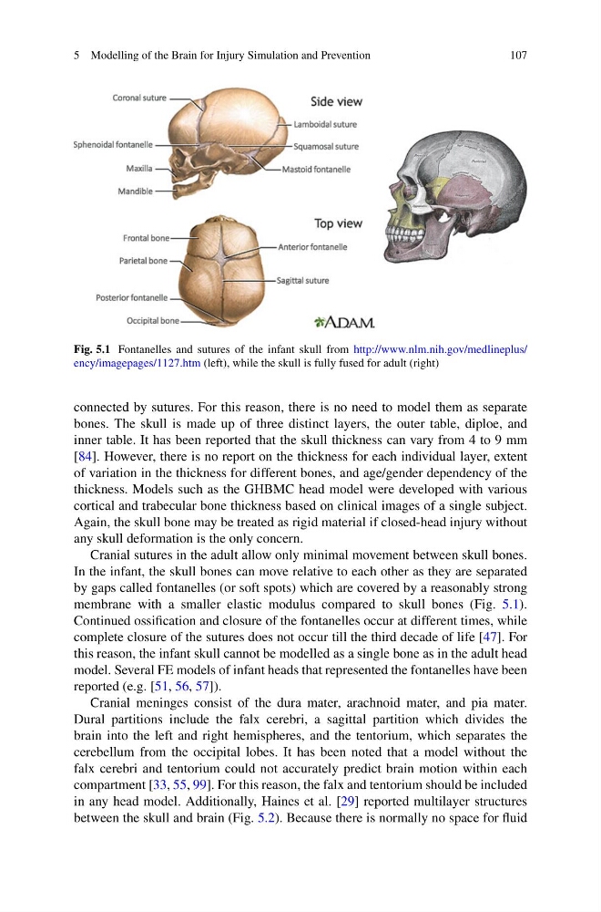
Fig. 5.1 Fontanelles and sutures of the infant skull from http://www.nlm.nih.gov/medlineplus/
ency/imagepages/1127.htm (left), while the skull is fully fused for adult (right)
connected by sutures. For this reason, there is no need to model them as separate
bones. The skull is made up of three distinct layers, the outer table, diploe, and
inner table. It has been reported that the skull thickness can vary from 4 to 9 mm
[84]. However, there is no report on the thickness for each individual layer, extent
of variation in the thickness for different bones, and age/gender dependency of the
thickness. Models such as the GHBMC head model were developed with various
cortical and trabecular bone thickness based on clinical images of a single subject.
Again, the skull bone may be treated as rigid material if closed-head injury without
any skull deformation is the only concern.
Cranial sutures in the adult allow only minimal movement between skull bones.
In the infant, the skull bones can move relative to each other as they are separated
by gaps called fontanelles (or soft spots) which are covered by a reasonably strong
membrane with a smaller elastic modulus compared to skull bones (Fig. 5.1).
Continued ossification and closure of the fontanelles occur at different times, while
complete closure of the sutures does not occur till the third decade of life [47]. For
this reason, the infant skull cannot be modelled as a single bone as in the adult head
model. Several FE models of infant heads that represented the fontanelles have been
reported (e.g. [51, 56, 57]).
Cranial meninges consist of the dura mater, arachnoid mater, and pia mater.
Dural partitions include the falx cerebri, a sagittal partition which divides the
brain into the left and right hemispheres, and the tentorium, which separates the
cerebellum from the occipital lobes. It has been noted that a model without the
falx cerebri and tentorium could not accurately predict brain motion within each
compartment [33, 55, 99]. For this reason, the falx and tentorium should be included
in any head model. Additionally, Haines et al. [29] reported multilayer structures
between the skull and brain (Fig. 5.2). Because there is normally no space for fluid
107
Fig. 5.1 Fontanelles and sutures of the infant skull from http://www.nlm.nih.gov/medlineplus/
ency/imagepages/1127.htm (left), while the skull is fully fused for adult (right)
connected by sutures. For this reason, there is no need to model them as separate
bones. The skull is made up of three distinct layers, the outer table, diploe, and
inner table. It has been reported that the skull thickness can vary from 4 to 9 mm
[84]. However, there is no report on the thickness for each individual layer, extent
of variation in the thickness for different bones, and age/gender dependency of the
thickness. Models such as the GHBMC head model were developed with various
cortical and trabecular bone thickness based on clinical images of a single subject.
Again, the skull bone may be treated as rigid material if closed-head injury without
any skull deformation is the only concern.
Cranial sutures in the adult allow only minimal movement between skull bones.
In the infant, the skull bones can move relative to each other as they are separated
by gaps called fontanelles (or soft spots) which are covered by a reasonably strong
membrane with a smaller elastic modulus compared to skull bones (Fig. 5.1).
Continued ossification and closure of the fontanelles occur at different times, while
complete closure of the sutures does not occur till the third decade of life [47]. For
this reason, the infant skull cannot be modelled as a single bone as in the adult head
model. Several FE models of infant heads that represented the fontanelles have been
reported (e.g. [51, 56, 57]).
Cranial meninges consist of the dura mater, arachnoid mater, and pia mater.
Dural partitions include the falx cerebri, a sagittal partition which divides the
brain into the left and right hemispheres, and the tentorium, which separates the
cerebellum from the occipital lobes. It has been noted that a model without the
falx cerebri and tentorium could not accurately predict brain motion within each
compartment [33, 55, 99]. For this reason, the falx and tentorium should be included
in any head model. Additionally, Haines et al. [29] reported multilayer structures
between the skull and brain (Fig. 5.2). Because there is normally no space for fluid