

58
3 Study of Electrophysical Characteristics of Blood …
540
550
560
570
580
590
600
610
620
630
640
1.24
1.242
1.244
1.246
1.248
1.25
1.252
1.254
1.256
1.258
x10
-4
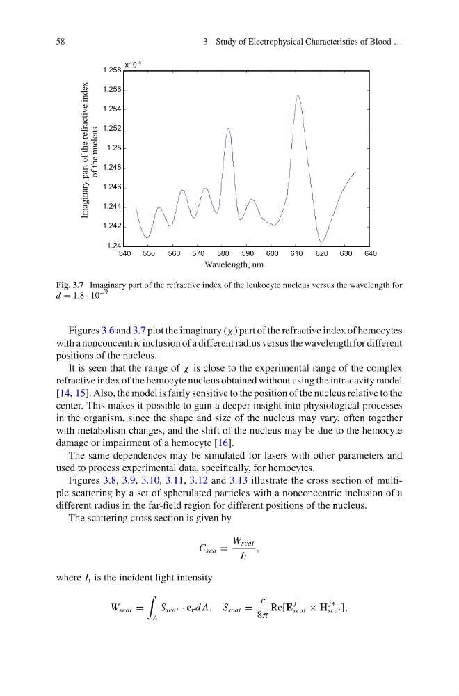
Fig. 3.7 Imaginary part of the refractive index of the leukocyte nucleus versus the wavelength for
d = 1.8 · 10 −7
Figures 3.6 and 3.7 plot the imaginary (χ ) part of the refractive index of hemocytes
with a nonconcentric inclusion of a different radius versus the wavelength for different
positions of the nucleus.
It is seen that the range of χ is close to the experimental range of the complex
refractive index of the hemocyte nucleus obtained without using the intracavity model
[14, 15]. Also, the model is fairly sensitive to the position of the nucleus relative to the
center. This makes it possible to gain a deeper insight into physiological processes
in the organism, since the shape and size of the nucleus may vary, often together
with metabolism changes, and the shift of the nucleus may be due to the hemocyte
damage or impairment of a hemocyte [16].
The same dependences may be simulated for lasers with other parameters and
used to process experimental data, specifically, for hemocytes.
Figures 3.8, 3.9, 3.10, 3.11, 3.12 and 3.13 illustrate the cross section of multiple scattering by a set of spherulated particles with a nonconcentric inclusion of a
different radius in the far-field region for different positions of the nucleus.
The scattering cross section is given by
C sca =
W scat
I i
,
where I i is the incident light intensity
W scat =
A
S scat · e r d A, S scat =
c
8π
Re[E
j
scat × H
j∗
scat ],
3 Study of Electrophysical Characteristics of Blood …
540
550
560
570
580
590
600
610
620
630
640
1.24
1.242
1.244
1.246
1.248
1.25
1.252
1.254
1.256
1.258
x10
-4
Fig. 3.7 Imaginary part of the refractive index of the leukocyte nucleus versus the wavelength for
d = 1.8 · 10 −7
Figures 3.6 and 3.7 plot the imaginary (χ ) part of the refractive index of hemocytes
with a nonconcentric inclusion of a different radius versus the wavelength for different
positions of the nucleus.
It is seen that the range of χ is close to the experimental range of the complex
refractive index of the hemocyte nucleus obtained without using the intracavity model
[14, 15]. Also, the model is fairly sensitive to the position of the nucleus relative to the
center. This makes it possible to gain a deeper insight into physiological processes
in the organism, since the shape and size of the nucleus may vary, often together
with metabolism changes, and the shift of the nucleus may be due to the hemocyte
damage or impairment of a hemocyte [16].
The same dependences may be simulated for lasers with other parameters and
used to process experimental data, specifically, for hemocytes.
Figures 3.8, 3.9, 3.10, 3.11, 3.12 and 3.13 illustrate the cross section of multiple scattering by a set of spherulated particles with a nonconcentric inclusion of a
different radius in the far-field region for different positions of the nucleus.
The scattering cross section is given by
C sca =
W scat
I i
,
where I i is the incident light intensity
W scat =
A
S scat · e r d A, S scat =
c
8π
Re[E
j
scat × H
j∗
scat ],