

158
9 Study of Optical Properties of Biotissues by the Intracavity …
(a)
(b)
nm
nm
10
-5
2 x
10
-5
2 x
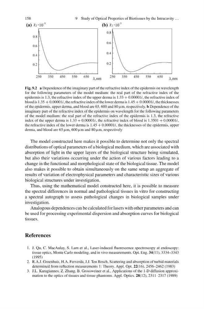
Fig. 9.2 a Dependence of the imaginary part of the refractive index of the epidermis on wavelength
for the following parameters of the model medium: the real part of the refractive index of the
epidermis is 1.3, the refractive index of the upper derma is 1.33 + 0.00001i, the refractive index of
blood is 1.35 + 0.00001i, the refractive index of the lower derma is 1.45 + 0.00001i, the thicknesses
of the epidermis, upper derma, and blood are 65, 600 and 80 µm, respectively. b Dependence of the
imaginary part of the refractive index of the epidermis on wavelength for the following parameters
of the model medium: the real part of the refractive index of the epidermis is 1.3, the refractive
index of the upper derma is 1.33 + 0.00001i, the refractive index of blood is 1.3501 + 0.00001i,
the refractive index of the lower derma is 1.45 + 0.00001i, the thicknesses of the epidermis, upper
derma, and blood are 65 µm, 600 µm and 80 µm, respectively
The model constructed here makes it possible to determine not only the spectral
distributions of optical parameters of a biological medium, which are associated with
absorption of light in the upper layers of the biological structure being simulated,
but also their variations occurring under the action of various factors leading to a
change in the functional and morphological state of the biological tissue. The model
also makes it possible to obtain simultaneously on the same setup an aggregate of
results of variation of electrophysical parameters and characteristic sizes of various
biological structures under investigation.
Thus, using the mathematical model constructed here, it is possible to measure
the spectral differences in normal and pathological tissues in vitro for constructing
a spectral autograph to assess pathological changes in biological samples under
investigation.
Analogous dependences can be calculated for lasers with other parameters and can
be used for processing experimental dispersion and absorption curves for biological
tissues.
References
1. J. Qu, C. MacAulay, S. Lam et al., Laser-induced fluorescence spectroscopy at endoscopy:
tissue optics, Monte Carlo modeling, and in vivo measurements. Opt. Eng. 34(11), 3334–3343
(1995)
2. R.A.J. Groenhuis, H.A. Ferverda, J.J. Ten Bosch, Scattering and absorption of turbid materials
determined from reflection measurements 1: Theory. Appl. Opt. 22(16), 2456–2462 (1983)
3. J.L. Karagiannes, Z. Zhang, B. Grossweiner et al., Applications of the 1-D diffusion approximation to the optics of tissues and tissue phantoms. Appl. Optics. 28(12), 2311–2317 (1989)
9 Study of Optical Properties of Biotissues by the Intracavity …
(a)
(b)
nm
nm
10
-5
2 x
10
-5
2 x
Fig. 9.2 a Dependence of the imaginary part of the refractive index of the epidermis on wavelength
for the following parameters of the model medium: the real part of the refractive index of the
epidermis is 1.3, the refractive index of the upper derma is 1.33 + 0.00001i, the refractive index of
blood is 1.35 + 0.00001i, the refractive index of the lower derma is 1.45 + 0.00001i, the thicknesses
of the epidermis, upper derma, and blood are 65, 600 and 80 µm, respectively. b Dependence of the
imaginary part of the refractive index of the epidermis on wavelength for the following parameters
of the model medium: the real part of the refractive index of the epidermis is 1.3, the refractive
index of the upper derma is 1.33 + 0.00001i, the refractive index of blood is 1.3501 + 0.00001i,
the refractive index of the lower derma is 1.45 + 0.00001i, the thicknesses of the epidermis, upper
derma, and blood are 65 µm, 600 µm and 80 µm, respectively
The model constructed here makes it possible to determine not only the spectral
distributions of optical parameters of a biological medium, which are associated with
absorption of light in the upper layers of the biological structure being simulated,
but also their variations occurring under the action of various factors leading to a
change in the functional and morphological state of the biological tissue. The model
also makes it possible to obtain simultaneously on the same setup an aggregate of
results of variation of electrophysical parameters and characteristic sizes of various
biological structures under investigation.
Thus, using the mathematical model constructed here, it is possible to measure
the spectral differences in normal and pathological tissues in vitro for constructing
a spectral autograph to assess pathological changes in biological samples under
investigation.
Analogous dependences can be calculated for lasers with other parameters and can
be used for processing experimental dispersion and absorption curves for biological
tissues.
References
1. J. Qu, C. MacAulay, S. Lam et al., Laser-induced fluorescence spectroscopy at endoscopy:
tissue optics, Monte Carlo modeling, and in vivo measurements. Opt. Eng. 34(11), 3334–3343
(1995)
2. R.A.J. Groenhuis, H.A. Ferverda, J.J. Ten Bosch, Scattering and absorption of turbid materials
determined from reflection measurements 1: Theory. Appl. Opt. 22(16), 2456–2462 (1983)
3. J.L. Karagiannes, Z. Zhang, B. Grossweiner et al., Applications of the 1-D diffusion approximation to the optics of tissues and tissue phantoms. Appl. Optics. 28(12), 2311–2317 (1989)