

90
Y. Zhang
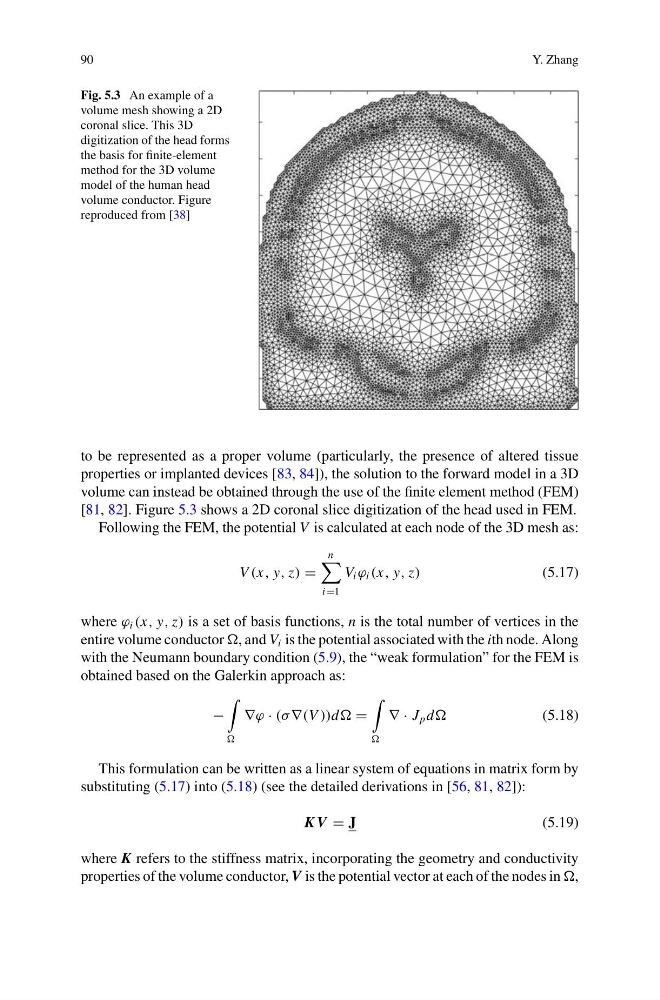
Fig. 5.3 An example of a
volume mesh showing a 2D
coronal slice. This 3D
digitization of the head forms
the basis for finite-element
method for the 3D volume
model of the human head
volume conductor. Figure
reproduced from [38]
to be represented as a proper volume (particularly, the presence of altered tissue
properties or implanted devices [83, 84]), the solution to the forward model in a 3D
volume can instead be obtained through the use of the finite element method (FEM)
[81, 82]. Figure 5.3 shows a 2D coronal slice digitization of the head used in FEM.
Following the FEM, the potential V is calculated at each node of the 3D mesh as:
V (x, y, z)
n
i1
V i ϕ i (x, y, z)
(5.17)
where ϕ i (x, y, z) is a set of basis functions, n is the total number of vertices in the
entire volume conductor , and V i is the potential associated with the ith node. Along
with the Neumann boundary condition (5.9), the “weak formulation” for the FEM is
obtained based on the Galerkin approach as:
−
∇ϕ · (σ ∇(V ))d
∇ · J p d
(5.18)
This formulation can be written as a linear system of equations in matrix form by
substituting (5.17) into (5.18) (see the detailed derivations in [56, 81, 82]):
K V J
(5.19)
where K refers to the stiffness matrix, incorporating the geometry and conductivity
properties of the volume conductor, V is the potential vector at each of the nodes in ,
Y. Zhang
Fig. 5.3 An example of a
volume mesh showing a 2D
coronal slice. This 3D
digitization of the head forms
the basis for finite-element
method for the 3D volume
model of the human head
volume conductor. Figure
reproduced from [38]
to be represented as a proper volume (particularly, the presence of altered tissue
properties or implanted devices [83, 84]), the solution to the forward model in a 3D
volume can instead be obtained through the use of the finite element method (FEM)
[81, 82]. Figure 5.3 shows a 2D coronal slice digitization of the head used in FEM.
Following the FEM, the potential V is calculated at each node of the 3D mesh as:
V (x, y, z)
n
i1
V i ϕ i (x, y, z)
(5.17)
where ϕ i (x, y, z) is a set of basis functions, n is the total number of vertices in the
entire volume conductor , and V i is the potential associated with the ith node. Along
with the Neumann boundary condition (5.9), the “weak formulation” for the FEM is
obtained based on the Galerkin approach as:
−
∇ϕ · (σ ∇(V ))d
∇ · J p d
(5.18)
This formulation can be written as a linear system of equations in matrix form by
substituting (5.17) into (5.18) (see the detailed derivations in [56, 81, 82]):
K V J
(5.19)
where K refers to the stiffness matrix, incorporating the geometry and conductivity
properties of the volume conductor, V is the potential vector at each of the nodes in ,