

8 Analysis of EEG in Medically Intractable Epilepsy
189
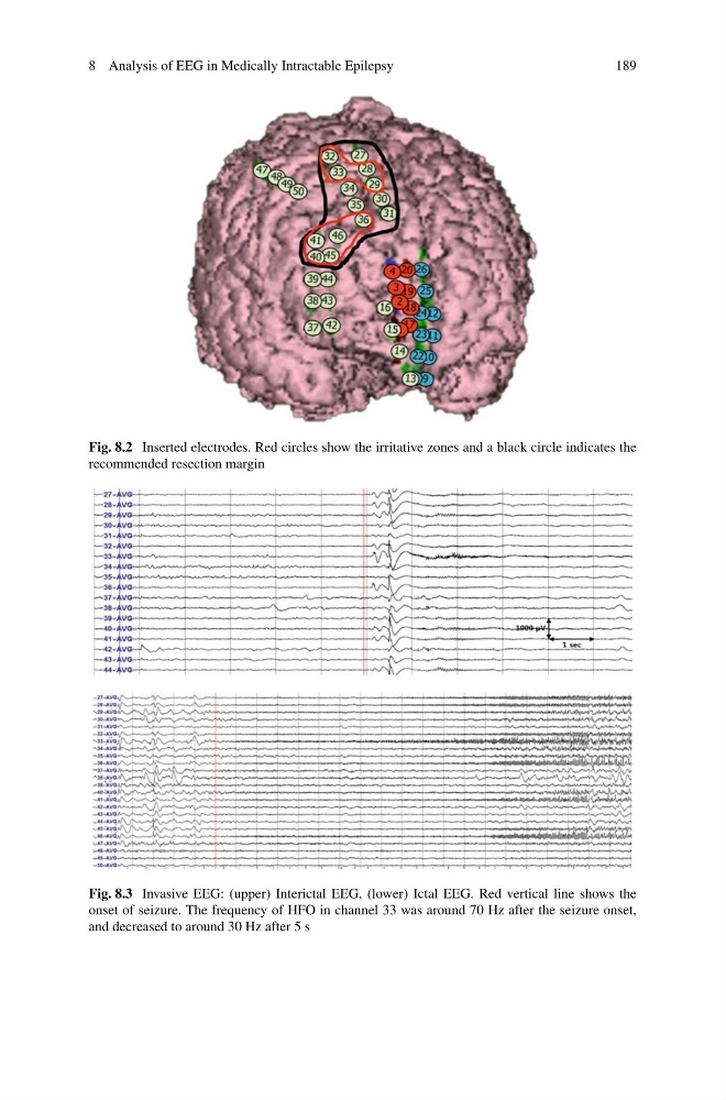
Fig. 8.2 Inserted electrodes. Red circles show the irritative zones and a black circle indicates the
recommended resection margin
Fig. 8.3 Invasive EEG: (upper) Interictal EEG, (lower) Ictal EEG. Red vertical line shows the
onset of seizure. The frequency of HFO in channel 33 was around 70 Hz after the seizure onset,
and decreased to around 30 Hz after 5 s
189
Fig. 8.2 Inserted electrodes. Red circles show the irritative zones and a black circle indicates the
recommended resection margin
Fig. 8.3 Invasive EEG: (upper) Interictal EEG, (lower) Ictal EEG. Red vertical line shows the
onset of seizure. The frequency of HFO in channel 33 was around 70 Hz after the seizure onset,
and decreased to around 30 Hz after 5 s