

188
K.-Y. Jung
sizes are significant but small. Therefore, randomized controlled clinical trials are
required to make convincing evidences in clinical practice.
8.3.5 Illustrated Case
This is a case of a 36-year-old man with epilepsy who showed the usefulness of HFO
in epilepsy surgery. He was right-handed and had no febrile convulsion episode,
and his seizures started at age of 22 years. The patient had no previous medical
history other than epilepsy. The patient had three types of seizure semiology: (1)
rotation rightward without impaired awareness and slightly impaired responsiveness,
(2) automotor or hypermotor seizure and (3) secondary generalized tonic-clonic
seizure. He had seizures two times a week while taking multiple AEDs including
carbamazepine 1200 mg, valproate 1200 mg, topiramate 200 mg and levetiracetam
1000 mg daily divided in two doses. Brain MRI, PET and interictal SPECT revealed
no definite abnormalities.
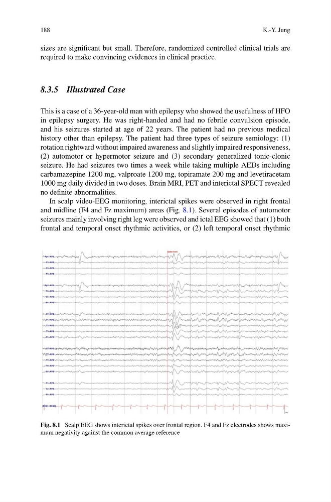
In scalp video-EEG monitoring, interictal spikes were observed in right frontal
and midline (F4 and Fz maximum) areas (Fig. 8.1). Several episodes of automotor
seizures mainly involving right leg were observed and ictal EEG showed that (1) both
frontal and temporal onset rhythmic activities, or (2) left temporal onset rhythmic
Fig. 8.1 Scalp EEG shows interictal spikes over frontal region. F4 and Fz electrodes shows maximum negativity against the common average reference
K.-Y. Jung
sizes are significant but small. Therefore, randomized controlled clinical trials are
required to make convincing evidences in clinical practice.
8.3.5 Illustrated Case
This is a case of a 36-year-old man with epilepsy who showed the usefulness of HFO
in epilepsy surgery. He was right-handed and had no febrile convulsion episode,
and his seizures started at age of 22 years. The patient had no previous medical
history other than epilepsy. The patient had three types of seizure semiology: (1)
rotation rightward without impaired awareness and slightly impaired responsiveness,
(2) automotor or hypermotor seizure and (3) secondary generalized tonic-clonic
seizure. He had seizures two times a week while taking multiple AEDs including
carbamazepine 1200 mg, valproate 1200 mg, topiramate 200 mg and levetiracetam
1000 mg daily divided in two doses. Brain MRI, PET and interictal SPECT revealed
no definite abnormalities.
In scalp video-EEG monitoring, interictal spikes were observed in right frontal
and midline (F4 and Fz maximum) areas (Fig. 8.1). Several episodes of automotor
seizures mainly involving right leg were observed and ictal EEG showed that (1) both
frontal and temporal onset rhythmic activities, or (2) left temporal onset rhythmic
Fig. 8.1 Scalp EEG shows interictal spikes over frontal region. F4 and Fz electrodes shows maximum negativity against the common average reference