

100
Y. Zhang
where f
ˆ
J
is the energy function associated with the prior, which can be interpreted
identically to the L
ˆ
J
term presented above.
Extending on this framework, Friston et al. introduced an EEG inverse method
called Multiple Sparse Priors (MSP) [27, 28] which formulates the forward problem
using a parametric empirical Bayesian approach on a hierarchical model [27–29].
MSP allows for the incorporation of multiple spatial covariance components into
the data model, weighted by a set of hyperparameters to be estimated empirically.
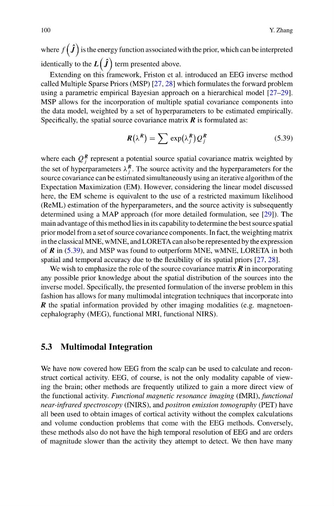
Specifically, the spatial source covariance matrix R is formulated as:
R
λ
R
exp
λ
R
j
Q
R
j
(5.39)
where each Q
R
j represent a potential source spatial covariance matrix weighted by
the set of hyperparameters λ
R
j . The source activity and the hyperparameters for the
source covariance can be estimated simultaneously using an iterative algorithm of the
Expectation Maximization (EM). However, considering the linear model discussed
here, the EM scheme is equivalent to the use of a restricted maximum likelihood
(ReML) estimation of the hyperparameters, and the source activity is subsequently
determined using a MAP approach (for more detailed formulation, see [29]). The
main advantage of this method lies in its capability to determine the best source spatial
prior model from a set of source covariance components. In fact, the weighting matrix
in the classical MNE, wMNE, and LORETA can also be represented by the expression
of R in (5.39), and MSP was found to outperform MNE, wMNE, LORETA in both
spatial and temporal accuracy due to the flexibility of its spatial priors [27, 28].
We wish to emphasize the role of the source covariance matrix R in incorporating
any possible prior knowledge about the spatial distribution of the sources into the
inverse model. Specifically, the presented formulation of the inverse problem in this
fashion has allows for many multimodal integration techniques that incorporate into
R the spatial information provided by other imaging modalities (e.g. magnetoencephalography (MEG), functional MRI, functional NIRS).
5.3 Multimodal Integration
We have now covered how EEG from the scalp can be used to calculate and reconstruct cortical activity. EEG, of course, is not the only modality capable of viewing the brain; other methods are frequently utilized to gain a more direct view of
the functional activity. Functional magnetic resonance imaging (fMRI), functional
near-infrared spectroscopy (fNIRS), and positron emission tomography (PET) have
all been used to obtain images of cortical activity without the complex calculations
and volume conduction problems that come with the EEG methods. Conversely,
these methods also do not have the high temporal resolution of EEG and are orders
of magnitude slower than the activity they attempt to detect. We then have many
Y. Zhang
where f
ˆ
J
is the energy function associated with the prior, which can be interpreted
identically to the L
ˆ
J
term presented above.
Extending on this framework, Friston et al. introduced an EEG inverse method
called Multiple Sparse Priors (MSP) [27, 28] which formulates the forward problem
using a parametric empirical Bayesian approach on a hierarchical model [27–29].
MSP allows for the incorporation of multiple spatial covariance components into
the data model, weighted by a set of hyperparameters to be estimated empirically.
Specifically, the spatial source covariance matrix R is formulated as:
R
λ
R
exp
λ
R
j
Q
R
j
(5.39)
where each Q
R
j represent a potential source spatial covariance matrix weighted by
the set of hyperparameters λ
R
j . The source activity and the hyperparameters for the
source covariance can be estimated simultaneously using an iterative algorithm of the
Expectation Maximization (EM). However, considering the linear model discussed
here, the EM scheme is equivalent to the use of a restricted maximum likelihood
(ReML) estimation of the hyperparameters, and the source activity is subsequently
determined using a MAP approach (for more detailed formulation, see [29]). The
main advantage of this method lies in its capability to determine the best source spatial
prior model from a set of source covariance components. In fact, the weighting matrix
in the classical MNE, wMNE, and LORETA can also be represented by the expression
of R in (5.39), and MSP was found to outperform MNE, wMNE, LORETA in both
spatial and temporal accuracy due to the flexibility of its spatial priors [27, 28].
We wish to emphasize the role of the source covariance matrix R in incorporating
any possible prior knowledge about the spatial distribution of the sources into the
inverse model. Specifically, the presented formulation of the inverse problem in this
fashion has allows for many multimodal integration techniques that incorporate into
R the spatial information provided by other imaging modalities (e.g. magnetoencephalography (MEG), functional MRI, functional NIRS).
5.3 Multimodal Integration
We have now covered how EEG from the scalp can be used to calculate and reconstruct cortical activity. EEG, of course, is not the only modality capable of viewing the brain; other methods are frequently utilized to gain a more direct view of
the functional activity. Functional magnetic resonance imaging (fMRI), functional
near-infrared spectroscopy (fNIRS), and positron emission tomography (PET) have
all been used to obtain images of cortical activity without the complex calculations
and volume conduction problems that come with the EEG methods. Conversely,
these methods also do not have the high temporal resolution of EEG and are orders
of magnitude slower than the activity they attempt to detect. We then have many