

2.7 Conclusion and Future Perspectives
In a broad sense, it is possible to ascertain the major role of nuclear molecular
imaging, specifically PET and SPECT, in the diagnosis and management of tumor
processes at the molecular and cellular levels. It also allows noninvasive visualization and monitoring of therapeutic responses and disease progression.
Furthermore, molecular imaging has the advantage of allowing in vivo characterization of the genetic changes involved oncogenesis, holding a predictive capability
for selection of optimal molecular therapeutic regimes, promising the best outcomes, on a per-case basis (i.e., personalized medicine). In both SPECT and PET,
the intensity of the imaging signal is proportional to the amount of the tracer and the
ability to image physiological and functional processes, guided by the pharmacokinetics and biodistribution of the radiotracer, which provides vital information
not available from conventional imaging techniques. However, insufficient signal
intensity, lack of stability, nonspecific interactions, and low circulation half-life are
still limitations that must be overcome. With that in mind, NPs may serve as the
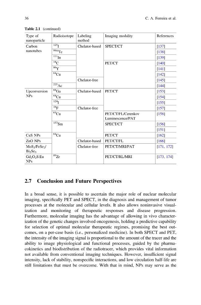
Table 2.1 (continued)
Type of
nanoparticle
Radioisotope Labeling
method
Imaging modality
References
Carbon
nanotubes
125
I
Chelator-based SPECT/CT
[137]
99m
Tc
[138]
111
In
[139]
14
C
PET/CT
[140]
86
Y
[ 141]
64
Cu
[142]
Chelator-free
[145]
225
Ac
[144]
Upconversion
NPs
68
Ga
Chelator-based PET/CT
[153]
64
Cu
[154]
124
I
[ 155]
18
F
Chelator-free
[157]
64
Cu
PET/CT/FL/Cerenkov
Luminescence/PAT
[158]
153
Sm
SPECT/CT
[156]
[151]
CuS NPs
64
Cu
PET/CT
[162]
ZnO NPs
Chelator-based PET/CT/FL
[166]
MoS 2 /FeSe 2 /
Bi 2 Se 3
Chelator-free
PET/CT/MRI/PAT
[171, 172]
Gd 2 O 2 S:Eu
NPs
89
Zr
PET/CT/RL/MRI
[173, 174]
36
C. A. Ferreira et al.
In a broad sense, it is possible to ascertain the major role of nuclear molecular
imaging, specifically PET and SPECT, in the diagnosis and management of tumor
processes at the molecular and cellular levels. It also allows noninvasive visualization and monitoring of therapeutic responses and disease progression.
Furthermore, molecular imaging has the advantage of allowing in vivo characterization of the genetic changes involved oncogenesis, holding a predictive capability
for selection of optimal molecular therapeutic regimes, promising the best outcomes, on a per-case basis (i.e., personalized medicine). In both SPECT and PET,
the intensity of the imaging signal is proportional to the amount of the tracer and the
ability to image physiological and functional processes, guided by the pharmacokinetics and biodistribution of the radiotracer, which provides vital information
not available from conventional imaging techniques. However, insufficient signal
intensity, lack of stability, nonspecific interactions, and low circulation half-life are
still limitations that must be overcome. With that in mind, NPs may serve as the
Table 2.1 (continued)
Type of
nanoparticle
Radioisotope Labeling
method
Imaging modality
References
Carbon
nanotubes
125
I
Chelator-based SPECT/CT
[137]
99m
Tc
[138]
111
In
[139]
14
C
PET/CT
[140]
86
Y
[ 141]
64
Cu
[142]
Chelator-free
[145]
225
Ac
[144]
Upconversion
NPs
68
Ga
Chelator-based PET/CT
[153]
64
Cu
[154]
124
I
[ 155]
18
F
Chelator-free
[157]
64
Cu
PET/CT/FL/Cerenkov
Luminescence/PAT
[158]
153
Sm
SPECT/CT
[156]
[151]
CuS NPs
64
Cu
PET/CT
[162]
ZnO NPs
Chelator-based PET/CT/FL
[166]
MoS 2 /FeSe 2 /
Bi 2 Se 3
Chelator-free
PET/CT/MRI/PAT
[171, 172]
Gd 2 O 2 S:Eu
NPs
89
Zr
PET/CT/RL/MRI
[173, 174]
36
C. A. Ferreira et al.