

and 16.2 ± 1.07%ID/g at 1, 24 and 48 h p.i.) [73]. Similarly, Zhou et al. [70]
demonstrated a one-step thermal reduction synthesis of NIR-emitting ultrasmall
radioactive glutathione (GS)-coated luminescent [
198 Au]Au NPs, with rapid and
effective renal clearance, as visualized through two different imaging modalities
(SPECT and optical imaging) [70].
As discussed in this section, a large number of studies support the use of
radioactive gold NPs as potential theranostic agents. However, a few limitations
regarding their use in biological applications have been found, such as low stability
in vivo [22] and lack of active tumor targeting [74]. In order to solve this problem,
PEG [74] coating of AuNPs and surface functionalization with different types of
biomolecules and ligands, such as peptides [75], DNA [76], RNA [77], antibodies
[78] has been extensively undertaken [74]. A thorough review covering all aspects
of bioconjugation of AuNP and their in vitro and in vivo characteristics can be
found elsewhere [79]. Perhaps the biggest challenge in using AuNPs for in vivo
purposes is their slow kinetics of elimination and considerable accumulation in the
liver and spleen. This potential concern with the use of AuNPs is evident
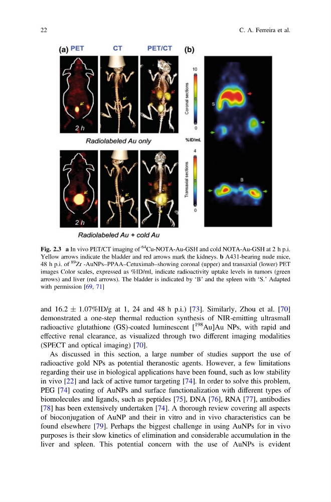
Fig. 2.3 a In vivo PET/CT imaging of
64 Cu-NOTA-Au-GSH and cold NOTA-Au-GSH at 2 h p.i.
Yellow arrows indicate the bladder and red arrows mark the kidneys. b A431-bearing nude mice,
48 h p.i. of
89
Zr -AuNPs–PPAA–Cetuximab–showing coronal (upper) and transaxial (lower) PET
images Color scales, expressed as %ID/ml, indicate radioactivity uptake levels in tumors (green
arrows) and liver (red arrows). The bladder is indicated by ‘B’ and the spleen with ‘S.’ Adapted
with permission [69, 71]
22
C. A. Ferreira et al.
demonstrated a one-step thermal reduction synthesis of NIR-emitting ultrasmall
radioactive glutathione (GS)-coated luminescent [
198 Au]Au NPs, with rapid and
effective renal clearance, as visualized through two different imaging modalities
(SPECT and optical imaging) [70].
As discussed in this section, a large number of studies support the use of
radioactive gold NPs as potential theranostic agents. However, a few limitations
regarding their use in biological applications have been found, such as low stability
in vivo [22] and lack of active tumor targeting [74]. In order to solve this problem,
PEG [74] coating of AuNPs and surface functionalization with different types of
biomolecules and ligands, such as peptides [75], DNA [76], RNA [77], antibodies
[78] has been extensively undertaken [74]. A thorough review covering all aspects
of bioconjugation of AuNP and their in vitro and in vivo characteristics can be
found elsewhere [79]. Perhaps the biggest challenge in using AuNPs for in vivo
purposes is their slow kinetics of elimination and considerable accumulation in the
liver and spleen. This potential concern with the use of AuNPs is evident
Fig. 2.3 a In vivo PET/CT imaging of
64 Cu-NOTA-Au-GSH and cold NOTA-Au-GSH at 2 h p.i.
Yellow arrows indicate the bladder and red arrows mark the kidneys. b A431-bearing nude mice,
48 h p.i. of
89
Zr -AuNPs–PPAA–Cetuximab–showing coronal (upper) and transaxial (lower) PET
images Color scales, expressed as %ID/ml, indicate radioactivity uptake levels in tumors (green
arrows) and liver (red arrows). The bladder is indicated by ‘B’ and the spleen with ‘S.’ Adapted
with permission [69, 71]
22
C. A. Ferreira et al.