

spectroscopy allowed the assessment of the breast cancer tissue margin after
operation; they also found and categorized cancer tissue which was ignored during
operation, important data to recommend additional treatment [50].
22.4 Molecular Imaging Using Radiolabeled NPs
22.4.1 SPECT
Gamma ray emissions are the source of SPECT images [51]. Radioisotopes decay
and emit gamma rays, which could be identified by a gamma camera to acquire
three-dimensional images. An emitted gamma photon meets collimator, which
permits gamma photons traveling along certain directions to correctly express the
source of gamma ray. Commonly used radioisotopes for SPECT are
99m Tc,
67 Ga,
111 In, and radioiodine (e.g.,
131 I) (Table 22.2).
The generator-produced SPECT radiotracer,
99m Tc, permits nuclear imaging
when cyclotron is not available.
99m
Tc (t1/2 = 6 h) permits SPECT imaging up to
1 day after injection, and it is the most commonly utilized radiotracer.
99m Tc has
been labeled to IONPs [52], silver NPs (AgNPs) [53], GNPs [54], and silica
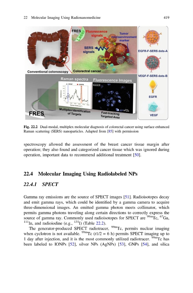
Fig. 22.2 Dual-modal, multiplex molecular diagnosis of colorectal cancer using surface enhanced
Raman scattering (SERS) nanoparticles. Adapted from [85] with permission
22 Molecular Imaging Using Radionanomedicine
419
operation; they also found and categorized cancer tissue which was ignored during
operation, important data to recommend additional treatment [50].
22.4 Molecular Imaging Using Radiolabeled NPs
22.4.1 SPECT
Gamma ray emissions are the source of SPECT images [51]. Radioisotopes decay
and emit gamma rays, which could be identified by a gamma camera to acquire
three-dimensional images. An emitted gamma photon meets collimator, which
permits gamma photons traveling along certain directions to correctly express the
source of gamma ray. Commonly used radioisotopes for SPECT are
99m Tc,
67 Ga,
111 In, and radioiodine (e.g.,
131 I) (Table 22.2).
The generator-produced SPECT radiotracer,
99m Tc, permits nuclear imaging
when cyclotron is not available.
99m
Tc (t1/2 = 6 h) permits SPECT imaging up to
1 day after injection, and it is the most commonly utilized radiotracer.
99m Tc has
been labeled to IONPs [52], silver NPs (AgNPs) [53], GNPs [54], and silica
Fig. 22.2 Dual-modal, multiplex molecular diagnosis of colorectal cancer using surface enhanced
Raman scattering (SERS) nanoparticles. Adapted from [85] with permission
22 Molecular Imaging Using Radionanomedicine
419