

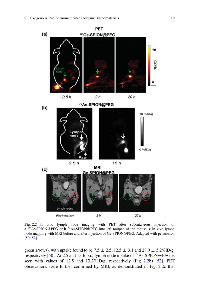
green arrows), with uptake found to be 7.5 ± 2.5, 12.5 ± 3.1 and 28.0 ± 5.2%ID/g,
respectively [50]. At 2.5 and 15 h p.i., lymph node uptake of
72 As-SPION@PEG is
seen with values of 12.5 and 13.2%ID/g, respectively (Fig. 2.2b) [52]. PET
observations were further confirmed by MRI, as demonstrated in Fig. 2.2c that
Fig. 2.2 In vivo lymph node imaging with PET after subcutaneous injection of
a
69
Ge-SPION@PEG or b
72
As-SPION@PEG into left footpad of the mouse. c In vivo lymph
node mapping with MRI before and after injection of Ge-SPION@PEG. Adapted with permission
[50, 52]
2 Exogenous Radionanomedicine: Inorganic Nanomaterials
19
respectively [50]. At 2.5 and 15 h p.i., lymph node uptake of
72 As-SPION@PEG is
seen with values of 12.5 and 13.2%ID/g, respectively (Fig. 2.2b) [52]. PET
observations were further confirmed by MRI, as demonstrated in Fig. 2.2c that
Fig. 2.2 In vivo lymph node imaging with PET after subcutaneous injection of
a
69
Ge-SPION@PEG or b
72
As-SPION@PEG into left footpad of the mouse. c In vivo lymph
node mapping with MRI before and after injection of Ge-SPION@PEG. Adapted with permission
[50, 52]
2 Exogenous Radionanomedicine: Inorganic Nanomaterials
19