

89 Zr through iron bonding and heat-induced methods. Thermodynamically and
kinetically stable
89
Zr–USPION were studied in vivo in a subcutaneous acute phase
response inflammation model, and results validated the potential of
89 Zr–USPION
as a PET/CT tracer of tissues enriched with activated macrophages. In the same
manner, through a chelator-free radiolabeling method, Cui et al. [51] took advantage of the high affinity between Al(OH) 3 and fluoride anions and labeled Al(OH) 3
coated IONPs with
18 F. High radiolabeling efficiency of 97% was achieved and,
after tail vein injection, a rapid accumulation of the radiolabeled IONPS was found
in the spleen and liver as confirmed by in vivo PET/CT and PET/MR imaging.
However, due to the unstable Al(OH) 3 shell, [
18 F]-fluoride was progressively
released from NPs in vivo, resulting in a considerable bone accumulation demonstrating suboptimal radiolabeling stability. Our group further demonstrated the
feasibility of chelator-free radiolabeling of SPIONs with Germanium-69 (
69 Ge) [50]
and Arsenic-72 (
72 As) [52]. Water-soluble poly(acrylic acid) (PAA) modified
PEGylated SPION was used for both studies. While the high affinity of germanium
for metal oxides was explored for the
69 Ge labeling,
72 As labeling involved
occupation of vacant FeO 4 tetrahedral sites in magnetite NPs by As
III O 3 trigonal
pyramids and As
V O 4 tetrahedra, yielding highly stable complexes in both scenarios.
PET/MRI lymph node mapping in vivo was demonstrated with
72 As-SPION and
69 Ge-SPION, as can be seen in Fig. 2.2. Accumulation of
69 Ge-SPION@PEG in
the popliteal lymph node is evident at 0.5, 2, and 20 h p.i. (Fig. 2.2a, marked by
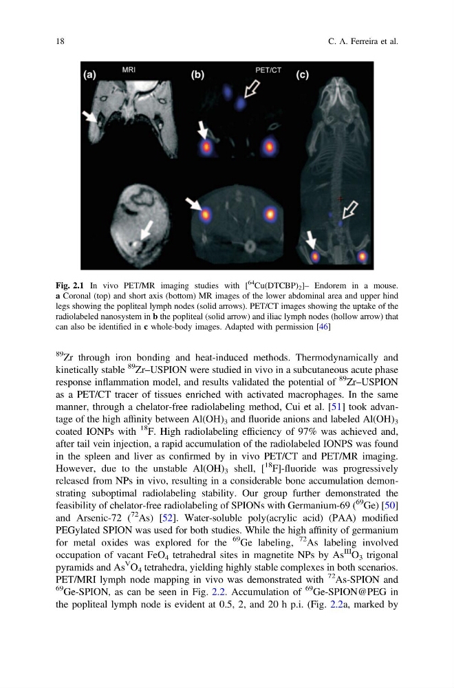
Fig. 2.1 In vivo PET/MR imaging studies with [
64
Cu(DTCBP) 2 ]– Endorem in a mouse.
a Coronal (top) and short axis (bottom) MR images of the lower abdominal area and upper hind
legs showing the popliteal lymph nodes (solid arrows). PET/CT images showing the uptake of the
radiolabeled nanosystem in b the popliteal (solid arrow) and iliac lymph nodes (hollow arrow) that
can also be identified in c whole-body images. Adapted with permission [46]
18
C. A. Ferreira et al.
kinetically stable
89
Zr–USPION were studied in vivo in a subcutaneous acute phase
response inflammation model, and results validated the potential of
89 Zr–USPION
as a PET/CT tracer of tissues enriched with activated macrophages. In the same
manner, through a chelator-free radiolabeling method, Cui et al. [51] took advantage of the high affinity between Al(OH) 3 and fluoride anions and labeled Al(OH) 3
coated IONPs with
18 F. High radiolabeling efficiency of 97% was achieved and,
after tail vein injection, a rapid accumulation of the radiolabeled IONPS was found
in the spleen and liver as confirmed by in vivo PET/CT and PET/MR imaging.
However, due to the unstable Al(OH) 3 shell, [
18 F]-fluoride was progressively
released from NPs in vivo, resulting in a considerable bone accumulation demonstrating suboptimal radiolabeling stability. Our group further demonstrated the
feasibility of chelator-free radiolabeling of SPIONs with Germanium-69 (
69 Ge) [50]
and Arsenic-72 (
72 As) [52]. Water-soluble poly(acrylic acid) (PAA) modified
PEGylated SPION was used for both studies. While the high affinity of germanium
for metal oxides was explored for the
69 Ge labeling,
72 As labeling involved
occupation of vacant FeO 4 tetrahedral sites in magnetite NPs by As
III O 3 trigonal
pyramids and As
V O 4 tetrahedra, yielding highly stable complexes in both scenarios.
PET/MRI lymph node mapping in vivo was demonstrated with
72 As-SPION and
69 Ge-SPION, as can be seen in Fig. 2.2. Accumulation of
69 Ge-SPION@PEG in
the popliteal lymph node is evident at 0.5, 2, and 20 h p.i. (Fig. 2.2a, marked by
Fig. 2.1 In vivo PET/MR imaging studies with [
64
Cu(DTCBP) 2 ]– Endorem in a mouse.
a Coronal (top) and short axis (bottom) MR images of the lower abdominal area and upper hind
legs showing the popliteal lymph nodes (solid arrows). PET/CT images showing the uptake of the
radiolabeled nanosystem in b the popliteal (solid arrow) and iliac lymph nodes (hollow arrow) that
can also be identified in c whole-body images. Adapted with permission [46]
18
C. A. Ferreira et al.