

(ABC). This is caused by the formation of anti-PEG IgM antibody after the first
injection of the PEGylated NPs. When the same PEGylated NPs are injected, the
preformed anti-PEG IgM recognizes the PEGylated NPs. Consequently,
opsonization and complement activation take place which can facilitate RES
recognition of the NPs [44]. Another way to reduce opsonization is called “active
stealth” approach. Rodriguez et al. attached CD47 ‘self’ peptides on the NPs, and
the NPs showed longer blood half-life by delaying splenic phagocytic clearance
[45]. Biomimetic particle coating can also reduce the opsonization and consequent
phagocytic clearance. Parodi et al. reported that NP coated using cell membranes
from leukocytes showed a reduction of serum protein attachment and lower MPS
uptake [46].
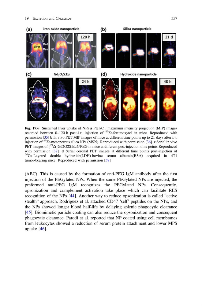
Fig. 19.6 Sustained liver uptake of NPs a PET/CT maximum intensity projection (MIP) images
recorded between 0–120 h post-i.v. injection of
89
Zr-ferumoxytol in mice. Reproduced with
permission [35] b In vivo PET MIP images of mice at different time points up to 21 days after i.v.
injection of
89
Zr-mesoporous silica NPs (MSN). Reproduced with permission [36]. c Serial in vivo
PET images of [
89
Zr]Gd2O2S:Eu@PEG in mice at different post-injection time points Reproduced
with permission [37]. d Serial coronal PET images at different time points post-injection of
64
Cu-Layered double hydroxide(LDH)-bovine serum albumin(BSA) acquired in 4T1
tumor-bearing mice. Reproduced with permission [38]
19 Excretion and Clearance
357
injection of the PEGylated NPs. When the same PEGylated NPs are injected, the
preformed anti-PEG IgM recognizes the PEGylated NPs. Consequently,
opsonization and complement activation take place which can facilitate RES
recognition of the NPs [44]. Another way to reduce opsonization is called “active
stealth” approach. Rodriguez et al. attached CD47 ‘self’ peptides on the NPs, and
the NPs showed longer blood half-life by delaying splenic phagocytic clearance
[45]. Biomimetic particle coating can also reduce the opsonization and consequent
phagocytic clearance. Parodi et al. reported that NP coated using cell membranes
from leukocytes showed a reduction of serum protein attachment and lower MPS
uptake [46].
Fig. 19.6 Sustained liver uptake of NPs a PET/CT maximum intensity projection (MIP) images
recorded between 0–120 h post-i.v. injection of
89
Zr-ferumoxytol in mice. Reproduced with
permission [35] b In vivo PET MIP images of mice at different time points up to 21 days after i.v.
injection of
89
Zr-mesoporous silica NPs (MSN). Reproduced with permission [36]. c Serial in vivo
PET images of [
89
Zr]Gd2O2S:Eu@PEG in mice at different post-injection time points Reproduced
with permission [37]. d Serial coronal PET images at different time points post-injection of
64
Cu-Layered double hydroxide(LDH)-bovine serum albumin(BSA) acquired in 4T1
tumor-bearing mice. Reproduced with permission [38]
19 Excretion and Clearance
357