

between renal clearance and blood circulation time. Only by addressing these
problems we can further enhance the in vivo tumor targeting efficacy of renal
clearable nanoparticles. For PK investigations of other radiolabeled nanoparticles,
such as iron oxide nanoparticle, upconversion nanoparticle, readers are advised to
refer to reviews or articles [89, 90].
17.3 Impact of Particle Shape and Surface Charge
Early work on polymer-based nanoparticles have shown the effect of nanoparticle’s
shape on the circulation time, efficacy of drug delivery as well as targeting efficacy
to the lungs and brain [91–93]. Although general conclusions about how to select
the optimal shape for certain applications could be drawn from previous review
articles [22, 94], it is still not clear how these could be applied to other nanoplatforms or in other animal models. So far, very few systematic studies were reported
on shape-dependent PK (or tumor-targeting) using radiolabeled nanoparticles.
Using intravital microscopy and QDs (HD size: 20–25 nm) and single-walled
carbon nanotubes (SWNTs) (*200 nm in length) with similar charge, surface
coating, and density as the model nanoparticles, researchers have shown a highly
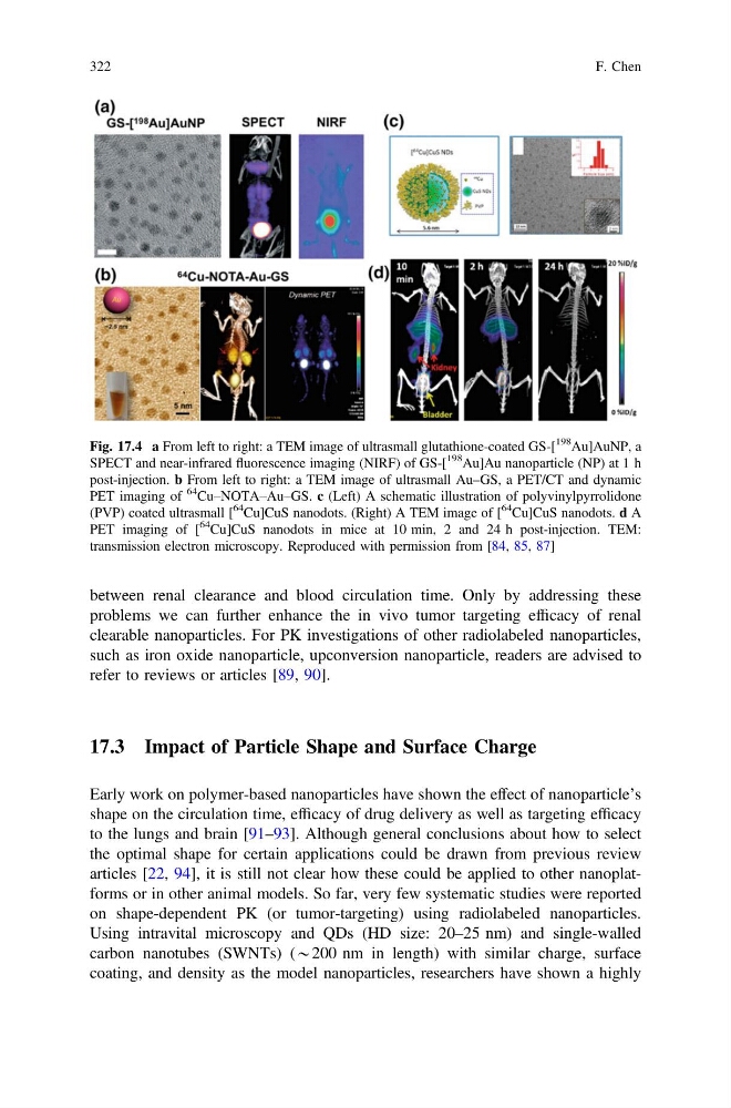
Fig. 17.4 a From left to right: a TEM image of ultrasmall glutathione-coated GS-[
198
Au]AuNP, a
SPECT and near-infrared fluorescence imaging (NIRF) of GS-[
198
Au]Au nanoparticle (NP) at 1 h
post-injection. b From left to right: a TEM image of ultrasmall Au–GS, a PET/CT and dynamic
PET imaging of
64
Cu–NOTA–Au–GS. c (Left) A schematic illustration of polyvinylpyrrolidone
(PVP) coated ultrasmall [
64
Cu]CuS nanodots. (Right) A TEM image of [
64
Cu]CuS nanodots. d A
PET imaging of [
64
Cu]CuS nanodots in mice at 10 min, 2 and 24 h post-injection. TEM:
transmission electron microscopy. Reproduced with permission from [84, 85, 87]
322
F. Chen
problems we can further enhance the in vivo tumor targeting efficacy of renal
clearable nanoparticles. For PK investigations of other radiolabeled nanoparticles,
such as iron oxide nanoparticle, upconversion nanoparticle, readers are advised to
refer to reviews or articles [89, 90].
17.3 Impact of Particle Shape and Surface Charge
Early work on polymer-based nanoparticles have shown the effect of nanoparticle’s
shape on the circulation time, efficacy of drug delivery as well as targeting efficacy
to the lungs and brain [91–93]. Although general conclusions about how to select
the optimal shape for certain applications could be drawn from previous review
articles [22, 94], it is still not clear how these could be applied to other nanoplatforms or in other animal models. So far, very few systematic studies were reported
on shape-dependent PK (or tumor-targeting) using radiolabeled nanoparticles.
Using intravital microscopy and QDs (HD size: 20–25 nm) and single-walled
carbon nanotubes (SWNTs) (*200 nm in length) with similar charge, surface
coating, and density as the model nanoparticles, researchers have shown a highly
Fig. 17.4 a From left to right: a TEM image of ultrasmall glutathione-coated GS-[
198
Au]AuNP, a
SPECT and near-infrared fluorescence imaging (NIRF) of GS-[
198
Au]Au nanoparticle (NP) at 1 h
post-injection. b From left to right: a TEM image of ultrasmall Au–GS, a PET/CT and dynamic
PET imaging of
64
Cu–NOTA–Au–GS. c (Left) A schematic illustration of polyvinylpyrrolidone
(PVP) coated ultrasmall [
64
Cu]CuS nanodots. (Right) A TEM image of [
64
Cu]CuS nanodots. d A
PET imaging of [
64
Cu]CuS nanodots in mice at 10 min, 2 and 24 h post-injection. TEM:
transmission electron microscopy. Reproduced with permission from [84, 85, 87]
322
F. Chen