

64 Cu [75]. It was found that radiolabeled QDs with a larger HD size had a faster
accumulation in both the liver and spleen, while PEGylated QDs exhibited slightly
slower rate of accumulation in these organs. The liver (27.4–38.9%ID/g) and spleen
(8.0–12.4%ID/g) uptake of
64 Cu-labeled QD800 (*21 nm) and QD525 (*12 nm)
were found to be quite close. A much higher liver (*50%ID/g) uptake was later
reported in RGD-functionalized,
64 Cu labeled QD705 in U87MG tumor-bearing
mice, possibly due to the larger particle size and relatively shorter blood circulation
half-life [76]. No evidence of QDs renal clearance was reported in either of these
early studies [75, 76].
A few months after this report, Choi et al. reported the first extensive
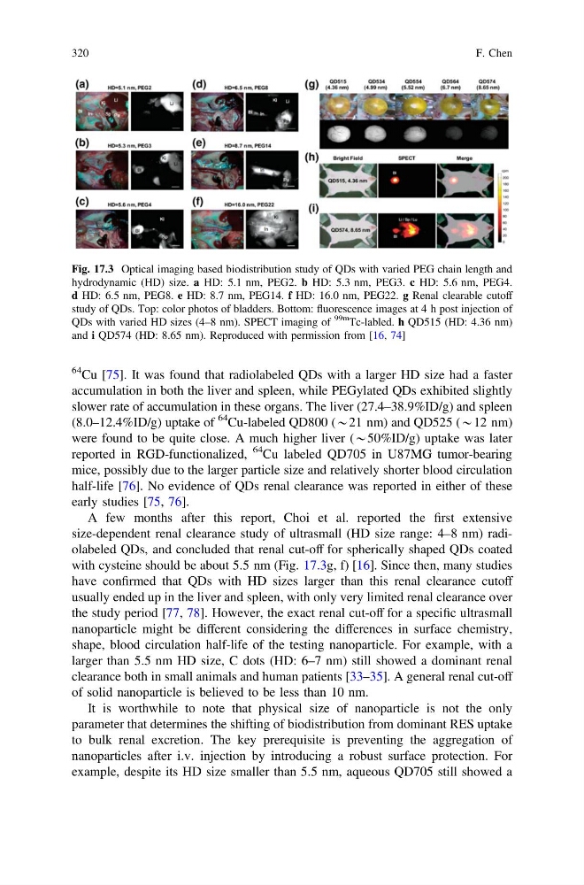
size-dependent renal clearance study of ultrasmall (HD size range: 4–8 nm) radiolabeled QDs, and concluded that renal cut-off for spherically shaped QDs coated
with cysteine should be about 5.5 nm (Fig. 17.3g, f) [16]. Since then, many studies
have confirmed that QDs with HD sizes larger than this renal clearance cutoff
usually ended up in the liver and spleen, with only very limited renal clearance over
the study period [77, 78]. However, the exact renal cut-off for a specific ultrasmall
nanoparticle might be different considering the differences in surface chemistry,
shape, blood circulation half-life of the testing nanoparticle. For example, with a
larger than 5.5 nm HD size, C dots (HD: 6–7 nm) still showed a dominant renal
clearance both in small animals and human patients [33–35]. A general renal cut-off
of solid nanoparticle is believed to be less than 10 nm.
It is worthwhile to note that physical size of nanoparticle is not the only
parameter that determines the shifting of biodistribution from dominant RES uptake
to bulk renal excretion. The key prerequisite is preventing the aggregation of
nanoparticles after i.v. injection by introducing a robust surface protection. For
example, despite its HD size smaller than 5.5 nm, aqueous QD705 still showed a
Fig. 17.3 Optical imaging based biodistribution study of QDs with varied PEG chain length and
hydrodynamic (HD) size. a HD: 5.1 nm, PEG2. b HD: 5.3 nm, PEG3. c HD: 5.6 nm, PEG4.
d HD: 6.5 nm, PEG8. e HD: 8.7 nm, PEG14. f HD: 16.0 nm, PEG22. g Renal clearable cutoff
study of QDs. Top: color photos of bladders. Bottom: fluorescence images at 4 h post injection of
QDs with varied HD sizes (4–8 nm). SPECT imaging of
99m
Tc-labled. h QD515 (HD: 4.36 nm)
and i QD574 (HD: 8.65 nm). Reproduced with permission from [16, 74]
320
F. Chen
accumulation in both the liver and spleen, while PEGylated QDs exhibited slightly
slower rate of accumulation in these organs. The liver (27.4–38.9%ID/g) and spleen
(8.0–12.4%ID/g) uptake of
64 Cu-labeled QD800 (*21 nm) and QD525 (*12 nm)
were found to be quite close. A much higher liver (*50%ID/g) uptake was later
reported in RGD-functionalized,
64 Cu labeled QD705 in U87MG tumor-bearing
mice, possibly due to the larger particle size and relatively shorter blood circulation
half-life [76]. No evidence of QDs renal clearance was reported in either of these
early studies [75, 76].
A few months after this report, Choi et al. reported the first extensive
size-dependent renal clearance study of ultrasmall (HD size range: 4–8 nm) radiolabeled QDs, and concluded that renal cut-off for spherically shaped QDs coated
with cysteine should be about 5.5 nm (Fig. 17.3g, f) [16]. Since then, many studies
have confirmed that QDs with HD sizes larger than this renal clearance cutoff
usually ended up in the liver and spleen, with only very limited renal clearance over
the study period [77, 78]. However, the exact renal cut-off for a specific ultrasmall
nanoparticle might be different considering the differences in surface chemistry,
shape, blood circulation half-life of the testing nanoparticle. For example, with a
larger than 5.5 nm HD size, C dots (HD: 6–7 nm) still showed a dominant renal
clearance both in small animals and human patients [33–35]. A general renal cut-off
of solid nanoparticle is believed to be less than 10 nm.
It is worthwhile to note that physical size of nanoparticle is not the only
parameter that determines the shifting of biodistribution from dominant RES uptake
to bulk renal excretion. The key prerequisite is preventing the aggregation of
nanoparticles after i.v. injection by introducing a robust surface protection. For
example, despite its HD size smaller than 5.5 nm, aqueous QD705 still showed a
Fig. 17.3 Optical imaging based biodistribution study of QDs with varied PEG chain length and
hydrodynamic (HD) size. a HD: 5.1 nm, PEG2. b HD: 5.3 nm, PEG3. c HD: 5.6 nm, PEG4.
d HD: 6.5 nm, PEG8. e HD: 8.7 nm, PEG14. f HD: 16.0 nm, PEG22. g Renal clearable cutoff
study of QDs. Top: color photos of bladders. Bottom: fluorescence images at 4 h post injection of
QDs with varied HD sizes (4–8 nm). SPECT imaging of
99m
Tc-labled. h QD515 (HD: 4.36 nm)
and i QD574 (HD: 8.65 nm). Reproduced with permission from [16, 74]
320
F. Chen