

With the advances of small animal SPECT, SPECT has a higher resolution than
that of PET unlikely with the clinical systems. Cheng et al. compared dedicated
small animal PET and SPECT systems [
99m Tc on a Nano-SPECT/CT camera
(Bioscan Inc., Washington D.C., USA) vs.
18 F on a Mosaic HP PET (Philips
Medical Systems, Inc., Cleveland, Ohio, USA)]. The phantom images and mouse
images using radiolabeled anti-HER2 NPs showed superior spatial resolution of
SPECT system (Fig. 15.6) [30].
Theranostic radioisotope including
177 Lu,
131 I can be utilized for both SPECT
imaging and radionuclide therapy.
177 Lu labeled lipid calcium phosphate
(LCP) NPs were used for tumor SPECT imaging and therapy. In a subcutaneous
xenograft tumor mouse model,
177 Lu–LCP showed the higher tumor uptake than
free
177 Lu. Also,
177 Lu–LCP demonstrated the superior ability to inhibit the tumor
growth than free
177 Lu [31]. SPECT/CT imaging was used to monitor the intratumorally injected
177 Lu labeled gold NPs in the subcutaneous breast cancer mouse
model. Regional distribution of radiation dose of a tumor can be calculated using
SPECT image. Finally, the NPs were able to inhibit the tumor growth and prolong
the survival (Fig. 15.7) [32]. Also, Ming et al. reported the development of theranostic
131 I labeled arginine-glycine-aspartate (RGD) bovine serum albumin
polycaprolactone for imaging and treatment of murine lung cancer models [33].
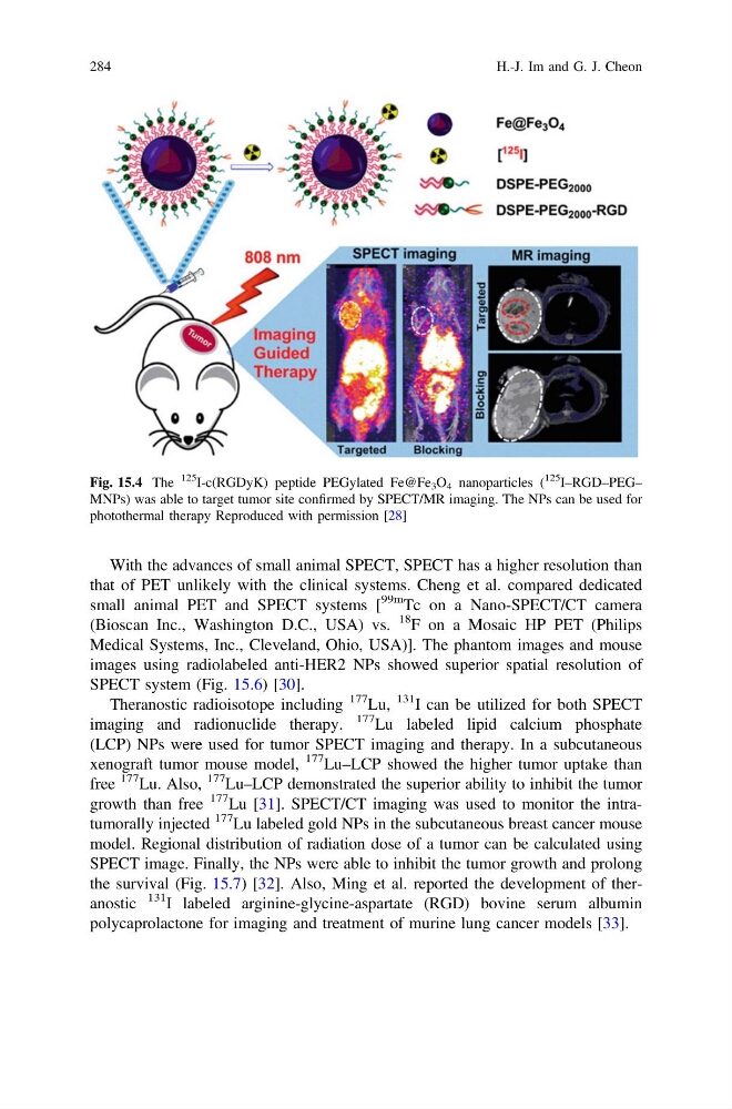
Fig. 15.4 The
125
I-c(RGDyK) peptide PEGylated Fe@Fe 3 O 4 nanoparticles (
125
I–RGD–PEG–
MNPs) was able to target tumor site confirmed by SPECT/MR imaging. The NPs can be used for
photothermal therapy Reproduced with permission [28]
284
H.-J. Im and G. J. Cheon
that of PET unlikely with the clinical systems. Cheng et al. compared dedicated
small animal PET and SPECT systems [
99m Tc on a Nano-SPECT/CT camera
(Bioscan Inc., Washington D.C., USA) vs.
18 F on a Mosaic HP PET (Philips
Medical Systems, Inc., Cleveland, Ohio, USA)]. The phantom images and mouse
images using radiolabeled anti-HER2 NPs showed superior spatial resolution of
SPECT system (Fig. 15.6) [30].
Theranostic radioisotope including
177 Lu,
131 I can be utilized for both SPECT
imaging and radionuclide therapy.
177 Lu labeled lipid calcium phosphate
(LCP) NPs were used for tumor SPECT imaging and therapy. In a subcutaneous
xenograft tumor mouse model,
177 Lu–LCP showed the higher tumor uptake than
free
177 Lu. Also,
177 Lu–LCP demonstrated the superior ability to inhibit the tumor
growth than free
177 Lu [31]. SPECT/CT imaging was used to monitor the intratumorally injected
177 Lu labeled gold NPs in the subcutaneous breast cancer mouse
model. Regional distribution of radiation dose of a tumor can be calculated using
SPECT image. Finally, the NPs were able to inhibit the tumor growth and prolong
the survival (Fig. 15.7) [32]. Also, Ming et al. reported the development of theranostic
131 I labeled arginine-glycine-aspartate (RGD) bovine serum albumin
polycaprolactone for imaging and treatment of murine lung cancer models [33].
Fig. 15.4 The
125
I-c(RGDyK) peptide PEGylated Fe@Fe 3 O 4 nanoparticles (
125
I–RGD–PEG–
MNPs) was able to target tumor site confirmed by SPECT/MR imaging. The NPs can be used for
photothermal therapy Reproduced with permission [28]
284
H.-J. Im and G. J. Cheon