

imaging. The NPs had 12 nm size and showed predominant splenic uptake (41.5%
ID/g) [24]. In vivo tracking of DNA NPs also have been reported using SPECT/CT
imaging. Patil et al. reported the time courses of
111 In labeled DNA micelle
deposition, and the distribution was compared between the different administration
routes, which are intrabiliary, hydrodynamic and intravenous infusion [25]. In
another study, SPECT/CT revealed the tumor targeting efficiency of
99m Tc labeled
tetrahedral DNA nanostructures [26].
There have been radionanoprobes for SPECT with the dual imaging ability.
99m Tc labeled superparamagnetic iron oxide (SPION) was developed and showed
dual imaging property for SPECT/MR for sentinel lymph node mapping [27].
Wang et al. developed a SPECT/MR probe,
125 I-c(RGDyK) peptide PEGylated
Fe@Fe 3 O 4 NPs (
125 I–RGD–PEG–MNPs). The probe was able to target glioblastoma xenograft tumor in the mouse model with the uptake of 6.75%ID/g, and the
uptake can be seen in both SPECT and MR images (Fig. 15.4) [28]. Black et al.
reported the development of dual radiolabeled (
125 I and
111 In) gold NPs. The single
SPECT scan can be separated into two SPECT images according to the energy
levels which showed distinct characteristics (Fig. 15.5) [29].
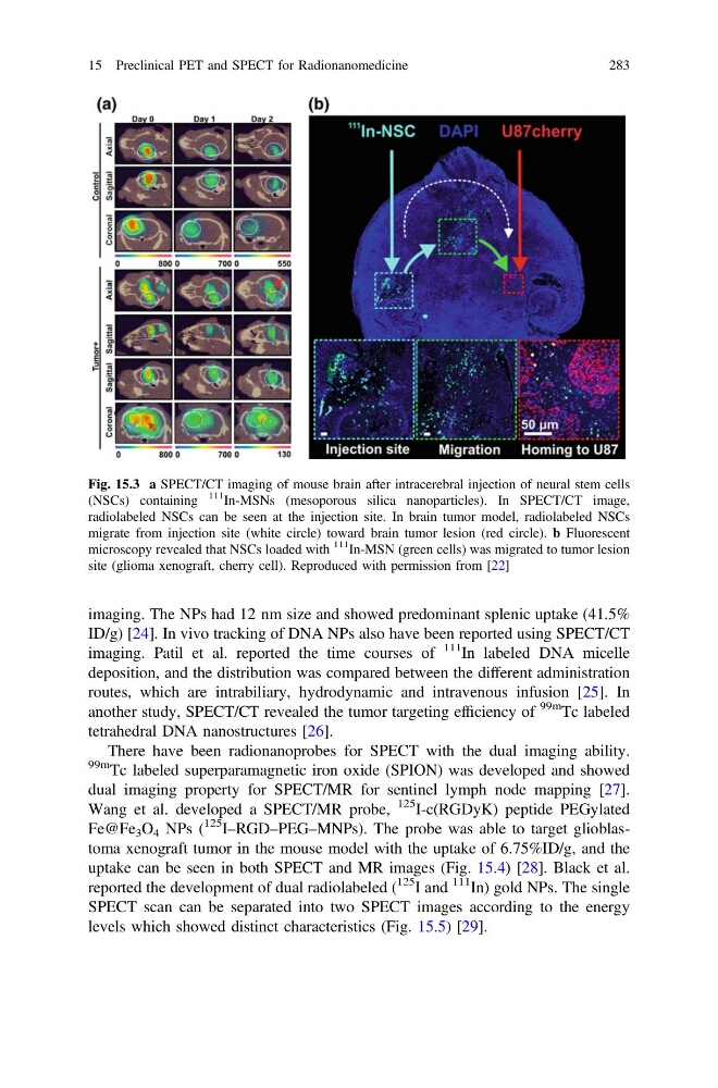
Fig. 15.3 a SPECT/CT imaging of mouse brain after intracerebral injection of neural stem cells
(NSCs) containing
111
In-MSNs (mesoporous silica nanoparticles). In SPECT/CT image,
radiolabeled NSCs can be seen at the injection site. In brain tumor model, radiolabeled NSCs
migrate from injection site (white circle) toward brain tumor lesion (red circle). b Fluorescent
microscopy revealed that NSCs loaded with
111
In-MSN (green cells) was migrated to tumor lesion
site (glioma xenograft, cherry cell). Reproduced with permission from [22]
15 Preclinical PET and SPECT for Radionanomedicine
283
ID/g) [24]. In vivo tracking of DNA NPs also have been reported using SPECT/CT
imaging. Patil et al. reported the time courses of
111 In labeled DNA micelle
deposition, and the distribution was compared between the different administration
routes, which are intrabiliary, hydrodynamic and intravenous infusion [25]. In
another study, SPECT/CT revealed the tumor targeting efficiency of
99m Tc labeled
tetrahedral DNA nanostructures [26].
There have been radionanoprobes for SPECT with the dual imaging ability.
99m Tc labeled superparamagnetic iron oxide (SPION) was developed and showed
dual imaging property for SPECT/MR for sentinel lymph node mapping [27].
Wang et al. developed a SPECT/MR probe,
125 I-c(RGDyK) peptide PEGylated
Fe@Fe 3 O 4 NPs (
125 I–RGD–PEG–MNPs). The probe was able to target glioblastoma xenograft tumor in the mouse model with the uptake of 6.75%ID/g, and the
uptake can be seen in both SPECT and MR images (Fig. 15.4) [28]. Black et al.
reported the development of dual radiolabeled (
125 I and
111 In) gold NPs. The single
SPECT scan can be separated into two SPECT images according to the energy
levels which showed distinct characteristics (Fig. 15.5) [29].
Fig. 15.3 a SPECT/CT imaging of mouse brain after intracerebral injection of neural stem cells
(NSCs) containing
111
In-MSNs (mesoporous silica nanoparticles). In SPECT/CT image,
radiolabeled NSCs can be seen at the injection site. In brain tumor model, radiolabeled NSCs
migrate from injection site (white circle) toward brain tumor lesion (red circle). b Fluorescent
microscopy revealed that NSCs loaded with
111
In-MSN (green cells) was migrated to tumor lesion
site (glioma xenograft, cherry cell). Reproduced with permission from [22]
15 Preclinical PET and SPECT for Radionanomedicine
283