

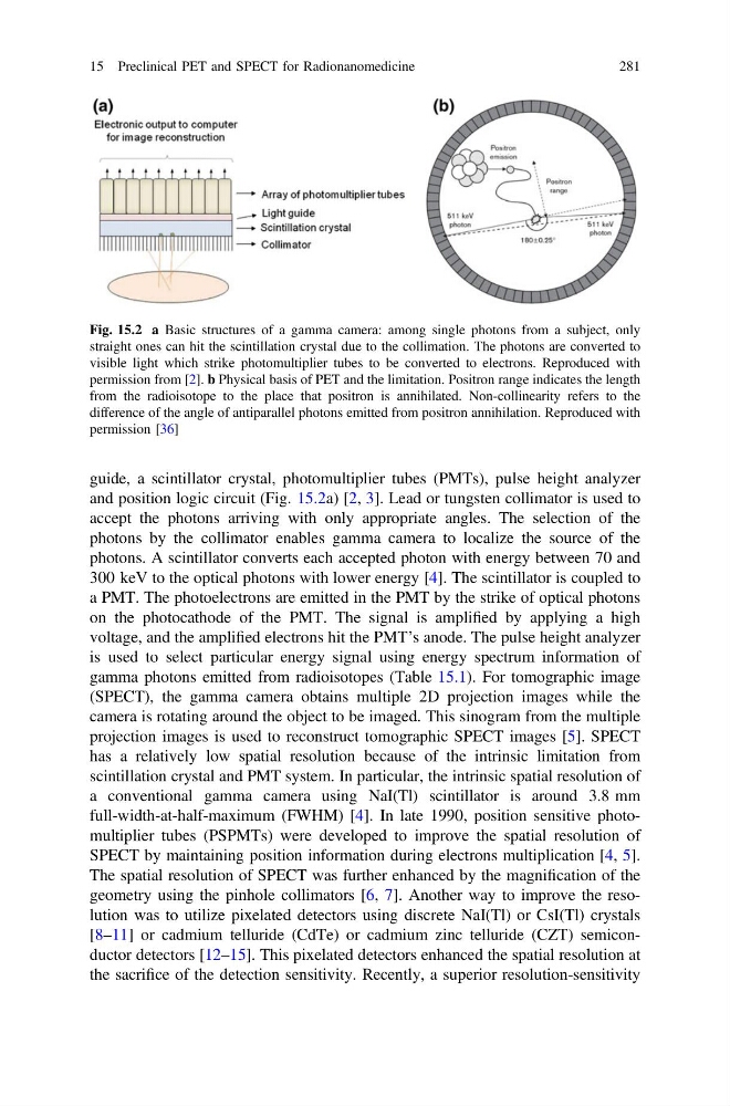
guide, a scintillator crystal, photomultiplier tubes (PMTs), pulse height analyzer
and position logic circuit (Fig. 15.2a) [2, 3]. Lead or tungsten collimator is used to
accept the photons arriving with only appropriate angles. The selection of the
photons by the collimator enables gamma camera to localize the source of the
photons. A scintillator converts each accepted photon with energy between 70 and
300 keV to the optical photons with lower energy [4]. The scintillator is coupled to
a PMT. The photoelectrons are emitted in the PMT by the strike of optical photons
on the photocathode of the PMT. The signal is amplified by applying a high
voltage, and the amplified electrons hit the PMT’s anode. The pulse height analyzer
is used to select particular energy signal using energy spectrum information of
gamma photons emitted from radioisotopes (Table 15.1). For tomographic image
(SPECT), the gamma camera obtains multiple 2D projection images while the
camera is rotating around the object to be imaged. This sinogram from the multiple
projection images is used to reconstruct tomographic SPECT images [5]. SPECT
has a relatively low spatial resolution because of the intrinsic limitation from
scintillation crystal and PMT system. In particular, the intrinsic spatial resolution of
a conventional gamma camera using NaI(Tl) scintillator is around 3.8 mm
full-width-at-half-maximum (FWHM) [4]. In late 1990, position sensitive photomultiplier tubes (PSPMTs) were developed to improve the spatial resolution of
SPECT by maintaining position information during electrons multiplication [4, 5].
The spatial resolution of SPECT was further enhanced by the magnification of the
geometry using the pinhole collimators [6, 7]. Another way to improve the resolution was to utilize pixelated detectors using discrete NaI(Tl) or CsI(Tl) crystals
[8–11] or cadmium telluride (CdTe) or cadmium zinc telluride (CZT) semiconductor detectors [12–15]. This pixelated detectors enhanced the spatial resolution at
the sacrifice of the detection sensitivity. Recently, a superior resolution-sensitivity
Fig. 15.2 a Basic structures of a gamma camera: among single photons from a subject, only
straight ones can hit the scintillation crystal due to the collimation. The photons are converted to
visible light which strike photomultiplier tubes to be converted to electrons. Reproduced with
permission from [2]. b Physical basis of PET and the limitation. Positron range indicates the length
from the radioisotope to the place that positron is annihilated. Non-collinearity refers to the
difference of the angle of antiparallel photons emitted from positron annihilation. Reproduced with
permission [36]
15 Preclinical PET and SPECT for Radionanomedicine
281
and position logic circuit (Fig. 15.2a) [2, 3]. Lead or tungsten collimator is used to
accept the photons arriving with only appropriate angles. The selection of the
photons by the collimator enables gamma camera to localize the source of the
photons. A scintillator converts each accepted photon with energy between 70 and
300 keV to the optical photons with lower energy [4]. The scintillator is coupled to
a PMT. The photoelectrons are emitted in the PMT by the strike of optical photons
on the photocathode of the PMT. The signal is amplified by applying a high
voltage, and the amplified electrons hit the PMT’s anode. The pulse height analyzer
is used to select particular energy signal using energy spectrum information of
gamma photons emitted from radioisotopes (Table 15.1). For tomographic image
(SPECT), the gamma camera obtains multiple 2D projection images while the
camera is rotating around the object to be imaged. This sinogram from the multiple
projection images is used to reconstruct tomographic SPECT images [5]. SPECT
has a relatively low spatial resolution because of the intrinsic limitation from
scintillation crystal and PMT system. In particular, the intrinsic spatial resolution of
a conventional gamma camera using NaI(Tl) scintillator is around 3.8 mm
full-width-at-half-maximum (FWHM) [4]. In late 1990, position sensitive photomultiplier tubes (PSPMTs) were developed to improve the spatial resolution of
SPECT by maintaining position information during electrons multiplication [4, 5].
The spatial resolution of SPECT was further enhanced by the magnification of the
geometry using the pinhole collimators [6, 7]. Another way to improve the resolution was to utilize pixelated detectors using discrete NaI(Tl) or CsI(Tl) crystals
[8–11] or cadmium telluride (CdTe) or cadmium zinc telluride (CZT) semiconductor detectors [12–15]. This pixelated detectors enhanced the spatial resolution at
the sacrifice of the detection sensitivity. Recently, a superior resolution-sensitivity
Fig. 15.2 a Basic structures of a gamma camera: among single photons from a subject, only
straight ones can hit the scintillation crystal due to the collimation. The photons are converted to
visible light which strike photomultiplier tubes to be converted to electrons. Reproduced with
permission from [2]. b Physical basis of PET and the limitation. Positron range indicates the length
from the radioisotope to the place that positron is annihilated. Non-collinearity refers to the
difference of the angle of antiparallel photons emitted from positron annihilation. Reproduced with
permission [36]
15 Preclinical PET and SPECT for Radionanomedicine
281