

the two tomographic imaging techniques which are used in radionanomedicine.
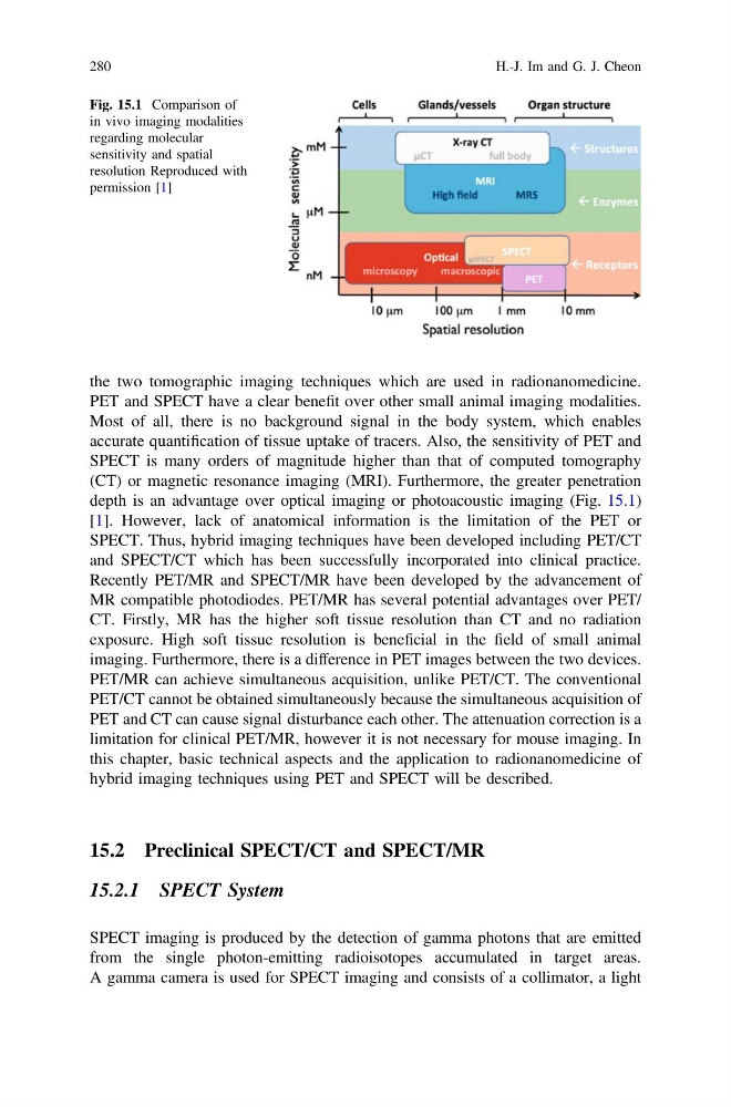
PET and SPECT have a clear benefit over other small animal imaging modalities.
Most of all, there is no background signal in the body system, which enables
accurate quantification of tissue uptake of tracers. Also, the sensitivity of PET and
SPECT is many orders of magnitude higher than that of computed tomography
(CT) or magnetic resonance imaging (MRI). Furthermore, the greater penetration
depth is an advantage over optical imaging or photoacoustic imaging (Fig. 15.1)
[1]. However, lack of anatomical information is the limitation of the PET or
SPECT. Thus, hybrid imaging techniques have been developed including PET/CT
and SPECT/CT which has been successfully incorporated into clinical practice.
Recently PET/MR and SPECT/MR have been developed by the advancement of
MR compatible photodiodes. PET/MR has several potential advantages over PET/
CT. Firstly, MR has the higher soft tissue resolution than CT and no radiation
exposure. High soft tissue resolution is beneficial in the field of small animal
imaging. Furthermore, there is a difference in PET images between the two devices.
PET/MR can achieve simultaneous acquisition, unlike PET/CT. The conventional
PET/CT cannot be obtained simultaneously because the simultaneous acquisition of
PET and CT can cause signal disturbance each other. The attenuation correction is a
limitation for clinical PET/MR, however it is not necessary for mouse imaging. In
this chapter, basic technical aspects and the application to radionanomedicine of
hybrid imaging techniques using PET and SPECT will be described.
15.2 Preclinical SPECT/CT and SPECT/MR
15.2.1 SPECT System
SPECT imaging is produced by the detection of gamma photons that are emitted
from the single photon-emitting radioisotopes accumulated in target areas.
A gamma camera is used for SPECT imaging and consists of a collimator, a light
Fig. 15.1 Comparison of
in vivo imaging modalities
regarding molecular
sensitivity and spatial
resolution Reproduced with
permission [1]
280
H.-J. Im and G. J. Cheon
PET and SPECT have a clear benefit over other small animal imaging modalities.
Most of all, there is no background signal in the body system, which enables
accurate quantification of tissue uptake of tracers. Also, the sensitivity of PET and
SPECT is many orders of magnitude higher than that of computed tomography
(CT) or magnetic resonance imaging (MRI). Furthermore, the greater penetration
depth is an advantage over optical imaging or photoacoustic imaging (Fig. 15.1)
[1]. However, lack of anatomical information is the limitation of the PET or
SPECT. Thus, hybrid imaging techniques have been developed including PET/CT
and SPECT/CT which has been successfully incorporated into clinical practice.
Recently PET/MR and SPECT/MR have been developed by the advancement of
MR compatible photodiodes. PET/MR has several potential advantages over PET/
CT. Firstly, MR has the higher soft tissue resolution than CT and no radiation
exposure. High soft tissue resolution is beneficial in the field of small animal
imaging. Furthermore, there is a difference in PET images between the two devices.
PET/MR can achieve simultaneous acquisition, unlike PET/CT. The conventional
PET/CT cannot be obtained simultaneously because the simultaneous acquisition of
PET and CT can cause signal disturbance each other. The attenuation correction is a
limitation for clinical PET/MR, however it is not necessary for mouse imaging. In
this chapter, basic technical aspects and the application to radionanomedicine of
hybrid imaging techniques using PET and SPECT will be described.
15.2 Preclinical SPECT/CT and SPECT/MR
15.2.1 SPECT System
SPECT imaging is produced by the detection of gamma photons that are emitted
from the single photon-emitting radioisotopes accumulated in target areas.
A gamma camera is used for SPECT imaging and consists of a collimator, a light
Fig. 15.1 Comparison of
in vivo imaging modalities
regarding molecular
sensitivity and spatial
resolution Reproduced with
permission [1]
280
H.-J. Im and G. J. Cheon