

consequently encapsulated within the nanoparticle framework. Following intravenous administration, these SNPs were found to accumulate within xenograft
tumours in mice as a result of the enhanced permeability and retention (EPR) effect
which is often the case for large macromolecular species [58–60]. Notably, these
particles then disassembled at the tumour site thereby affording access to the TCO
groups by a [
64 Cu]-radiolabeled Tz secondary agent. Promisingly, this pretargeting
approach resulted in the acquisition of PET images in which tumours were strongly
contrasted against surrounding tissues and organs.
In another notable example, Keinänen et al. have recently developed a pretargeting strategy based upon TCO-modified mesoporous silica nanoparticles and an
[
18 F]-labeled Tz secondary agent [61]. Mesoporous silica nanoparticles are an
attractive nanoplatform for this application due to their high loading potential, low
toxicity, and biodegradability. In this example, no tumour model was employed,
however the authors found evidence of IEDDA reaction products within the spleen.
While this particular approach did not involve targeting of any specific disease
biomarker, the promising ability of this reaction to proceed efficiently in vivo is a
good indicator of the potential that nanoparticle frameworks have to offer pretargeting applications in imaging and therapy.
In summary, click chemistry reactions have found great utility in the production
of a wide variety of radiopharmaceutical agents, particularly for PET radiotracers.
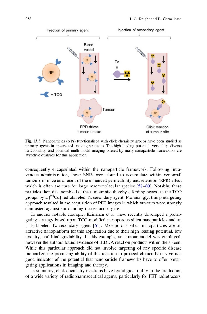
Fig. 13.5 Nanoparticles (NPs) functionalised with click chemistry groups have been studied as
primary agents in pretargeted imaging strategies. The high loading potential, versatility, diverse
functionality, and potential multi-modal imaging offered by many nanoparticle frameworks are
attractive qualities for this application
258
J. C. Knight and B. Cornelissen
tumours in mice as a result of the enhanced permeability and retention (EPR) effect
which is often the case for large macromolecular species [58–60]. Notably, these
particles then disassembled at the tumour site thereby affording access to the TCO
groups by a [
64 Cu]-radiolabeled Tz secondary agent. Promisingly, this pretargeting
approach resulted in the acquisition of PET images in which tumours were strongly
contrasted against surrounding tissues and organs.
In another notable example, Keinänen et al. have recently developed a pretargeting strategy based upon TCO-modified mesoporous silica nanoparticles and an
[
18 F]-labeled Tz secondary agent [61]. Mesoporous silica nanoparticles are an
attractive nanoplatform for this application due to their high loading potential, low
toxicity, and biodegradability. In this example, no tumour model was employed,
however the authors found evidence of IEDDA reaction products within the spleen.
While this particular approach did not involve targeting of any specific disease
biomarker, the promising ability of this reaction to proceed efficiently in vivo is a
good indicator of the potential that nanoparticle frameworks have to offer pretargeting applications in imaging and therapy.
In summary, click chemistry reactions have found great utility in the production
of a wide variety of radiopharmaceutical agents, particularly for PET radiotracers.
Fig. 13.5 Nanoparticles (NPs) functionalised with click chemistry groups have been studied as
primary agents in pretargeted imaging strategies. The high loading potential, versatility, diverse
functionality, and potential multi-modal imaging offered by many nanoparticle frameworks are
attractive qualities for this application
258
J. C. Knight and B. Cornelissen